TopPodcast.com
Menu
  • Home
  • Top Charts
  • Top Networks
  • Top Apps
  • Top Independents
  • Top Podfluencers
  • Top Picks
    • Top Business Podcasts
    • Top True Crime Podcasts
    • Top Finance Podcasts
    • Top Comedy Podcasts
    • Top Music Podcasts
    • Top Womens Podcasts
    • Top Kids Podcasts
    • Top Sports Podcasts
    • Top News Podcasts
    • Top Tech Podcasts
    • Top Crypto Podcasts
    • Top Entrepreneurial Podcasts
    • Top Fantasy Sports Podcasts
    • Top Political Podcasts
    • Top Science Podcasts
    • Top Self Help Podcasts
    • Top Sports Betting Podcasts
    • Top Stocks Podcasts
  • Podcast News
  • About Us
  • Podcast Advertising
  • Contact
Not in our directory?
Add Show Here
Podcast Equipment
Center

toppodcastlogoOur TOPPODCAST Picks

  • Comedy
  • Crypto
  • Sports
  • News
  • Politics
  • True Crime
  • Business
  • Finance

Follow Us

toppodcastlogoStay Connected

    View Top 200 Chart
    Back to Rankings Page
    Government & Organizations

    Core EM – Emergency Medicine Podcast

    Core EM Emergency Medicine Podcast

    Advertise

    Copyright: © Core EM

    • Apple Podcasts
    • Google Play
    • Spotify

    Latest Episodes:
    Episode 197: Acute Agitation Jun 03, 2024
    Show notes

    We discuss an approach to the acutely agitated patient and review medications commonly used.

    Hosts:
    Jonathan Kobles, MD
    Brian Gilberti, MD

    https://media.blubrry.com/coreem/content.blubrry.com/coreem/Acute_Agitation.mp3 Download Leave a Comment Tags: Agitation, psychiatry, Toxicology

    Show Notes

    Background/Epidemiology

    •Definition and Scope: Agitation encompasses behaviors from restlessness to severe altered mental states. It’s a common emergency department presentation, often linked with acute medical or psychiatric emergencies.

    •Significance: Patients with agitation are at high risk for morbidity and mortality, necessitating prompt and effective management to prevent harm to themselves and healthcare providers.

    A Changing Paradigm in Describing Agitation

    •Terminology Shift: Move away from terms like ‘excited delirium’ due to their politicization and stigmatization. Focus on describing agitation by severity and underlying causes.

    Agitation as a Multifactorial Process

    •Complex Nature: Recognize agitation as a result of various factors, including medical, psychiatric, and environmental influences.

    Recognizing Agitation

    •Signs and Symptoms: Identify agitation early by monitoring for behaviors such as hostility, pacing, non-compliance, and verbal aggression.

    Initial Evaluation

    •Severity Assessment: Determine the severity of agitation and prioritize reversible causes and life-threatening conditions.

    •Diagnostic Steps: Perform vital signs check, blood glucose levels, ECG, and a targeted medical screening exam.

    Life Threats

    •Immediate Concerns: Identify and address immediate life threats such as hypoxia, hypoglycemia, trauma, and acute neurological emergencies.

    Forming a Differential Prior to Treatment

    •Prioritization: Severe agitation requires immediate treatment to facilitate further evaluation and reduce risk of harm.

    Physician/Staff Safety

    •Safety Measures: Ensure personal and team safety by maintaining a calm environment and preparing for potential violence.

    Multimodal Approach

    •Self-check In: Physicians should mentally prepare and approach the situation calmly to ensure effective management.

    •Verbal De-escalation: Use techniques focused on safety, therapeutic alliance, and patient autonomy to manage agitation non-pharmacologically.

    Medication Administration

    •Oral/Sublingual Medications: Consider oral medications for less severe cases to maintain patient autonomy and avoid invasive procedures.

    •IM or IV Medications: Use intramuscular or intravenous medications for rapid control in severe cases.

    Specific Medication Regimens

    •PO Regimens:

    •Medications: Antipsychotics like Zyprexa (olanzapine) 5-10 mg, benzodiazepines like Ativan (lorazepam) 1-2 mg.

    •Benefits: Empower patients with a sense of autonomy, avoid injection-related trauma.

    •Pharmacokinetics:

    •Olanzapine: Onset in 15-45 minutes, peak effect in 1-2 hours, duration 12-24 hours.

    •Lorazepam: Onset in 30-60 minutes, peak effect in 2 hours, duration 6-8 hours.

    •IV/IM Regimens:

    •Medications: Droperidol, haloperidol, midazolam, ketamine.

    •ACEP 2023 Guidelines: Recommend droperidol with midazolam or an atypical antipsychotic for severe agitation.

    •Pharmacokinetics (IM):

    •Haloperidol: IM onset in 15, time to sedation ~25 minutes, can last for 2 hours

    •Droperidol: IM onset in 5-10 minutes, duration 2-4 hours but can last as long as 12 hours

    •Midazolam: IM onset ~15 minutes, , duration 20 minutes – 2 hours.

    •Lorazepam: IM onset ~15-30 minutes, , duration up to 3 hours

    •Ketamine: IM onset in ~5 minutes, duration 5-30 minutes.

    Special Situations

    •Elderly/Dementia: Optimize environment, use non-pharmacologic measures, avoid benzodiazepines to reduce delirium risk.

    •Parkinson’s Disease: Avoid antipsychotics that can precipitate a Parkinsonian crisis.

    •Autism/Pediatrics: Engage caregivers, create a calming environment, avoid aggressive measures.

    •Alcohol Withdrawal: Utilize benzodiazepines and phenobarbital.

    Re-dosing and Physical Restraints

    •Re-dosing: Use the lowest effective dose, consider continuous monitoring, and reassess frequently.

    •Physical Restraints: Employ as a last resort, ensuring close monitoring for any adverse effects.

    Final Points

    •Clinical Leadership: Physicians should lead with clear communication, planning, and support for the team.

    •Continuous Learning: Regular debriefing and assessment after each incident to improve future responses.


    Read More

    Episode 196: The Critically Ill Infant May 01, 2024
    Show notes

    We discuss an approach to the critically ill infant.

    Hosts:
    Ellen Duncan, MD, PhD
    Brian Gilberti, MD

    https://media.blubrry.com/coreem/content.blubrry.com/coreem/The_Critically_Ill_Infant.mp3 Download Leave a Comment Tags: Pediatrics

    Show Notes

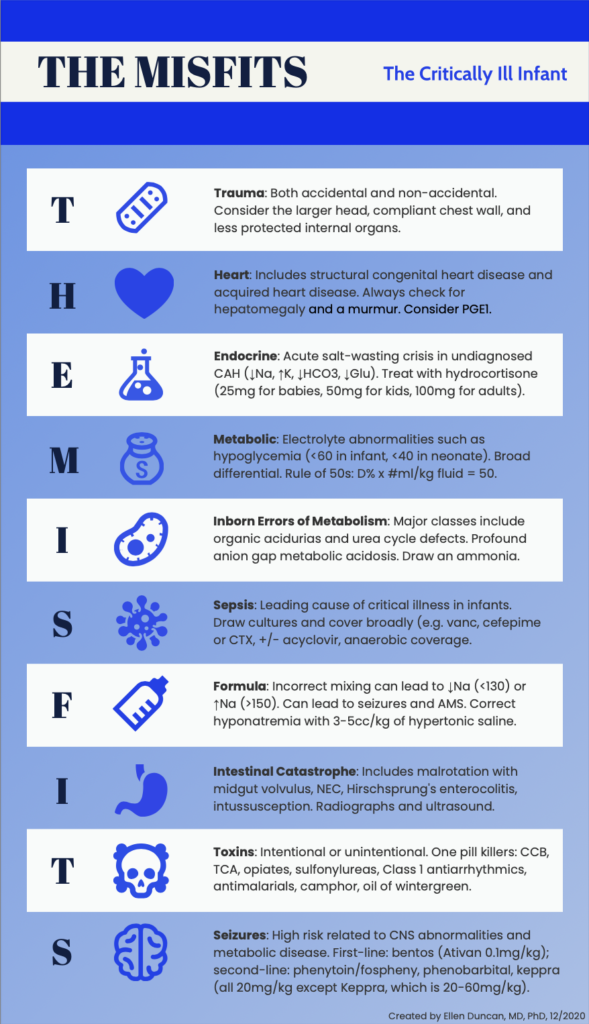
    The Critically Ill Infant: THE MISFITS

    Trauma

    • ‘T’ in the mnemonic stands for trauma, which includes both accidental and intentional causes.
    • Considerations for Non-accidental Trauma:
      • Stresses the importance of considering non-accidental trauma, especially given that it may not always present with obvious external signs.
    • Anatomical Vulnerabilities:
      • Highlights specific anatomical considerations for infants who suffer from trauma:
        • Infants have proportionally larger heads, increasing their susceptibility to high cervical spine (c-spine) injuries.
        • Their liver and spleen are less protected, making abdominal injuries potentially more severe.

    Heart

    • 5 T’s of Cyanotic Congenital Heart Disease: Introduces a mnemonic to help remember key right-sided ductal-dependent lesions:
      • Truncus Arteriosus: Single vessel serving as both pulmonary and systemic outflow tract.
      • Transposition of the Great Arteries: The pulmonary artery and aorta are switched, leading to improper circulation.
      • Tricuspid Atresia: Absence of the tricuspid valve, leading to inadequate development of the right ventricle and pulmonary circulation issues.
      • Tetralogy of Fallot: Comprises four defects—ventricular septal defect, pulmonary stenosis, right ventricular hypertrophy, and an overriding aorta.
      • Total Anomalous Pulmonary Venous Connection (TAPVC): Pulmonary veins do not connect to the left atrium but rather to the right heart or veins, causing oxygen-rich blood to mix with oxygen-poor blood.
    • Other Significant Conditions:
      • Ebstein’s Anomaly: Malformation of the tricuspid valve affecting right-sided heart function.
      • Pulmonary Atresia/Stenosis: Incomplete formation or narrowing of the pulmonary valve obstructs blood flow to the lungs.
    • Left-sided Ductal-Dependent Lesions:
      • Conditions such as aortic arch abnormalities (coarctation or interrupted arch), critical aortic stenosis, and hypoplastic left heart syndrome are highlighted. These generally present with less obvious cyanosis and more pallor.
    • Diagnostic and Management Considerations:
      • Routine prenatal ultrasounds detect most cases, but conditions like coarctation of the aorta and TAPVC might not be apparent until after birth when the ductus arteriosus closes.
      • Emphasizes the importance of a thorough physical exam: checking for murmurs, assessing hepatosplenomegaly, feeling for femoral pulses, measuring pre- and post-ductal saturations, and taking blood pressures in all four limbs.
    • Treatment Recommendations:
      • Early initiation of alprostadil (a prostaglandin) for patients with suspected ductal-dependent lesions to maintain ductal patency.
      • Preparedness for potential complications from alprostadil treatment, such as apnea and hypotension, which may necessitate intubation and hemodynamic support.

    Endocrine

    • Focuses on acute salt-wasting crisis in undiagnosed Congenital Adrenal Hyperplasia (CAH).
    • Electrolyte imbalances: ↓Na, ↑K, ↓HCO3, ↓Glu.
    • Treatment: hydrocortisone (25mg for babies, 50mg for kids, 100mg for adults).

    Metabolic

    • Electrolyte abnormalities such as hypoglycemia (values: <60 in infants, <40 in neonates).
    • Broad differential.
    • Rule of 50s for correction: D% x #ml/kg fluid = 50.

    Inborn Errors of Metabolism

    • Major classes include organic acidurias (profound anion gap metabolic acidosis) and urea cycle defects (hyperammonemia)
    • Recommendation: Draw gas and ammonia level.

    Sepsis

    • Emphasized as a critical condition in the differential diagnosis for ill infants, though placed later in the mnemonic for easier recall.
    • Presentation and Diagnosis:
      • Sepsis in infants often presents nonspecifically, making early detection challenging.
      • Immediate drawing of blood cultures upon suspicion of sepsis.
    • Initial Treatment:
      • Prompt initiation of antimicrobials and fluids.
      • Use of vancomycin for gram-positive and MRSA coverage, a third-generation cephalosporin or pip-tazo for broad bacterial coverage, and acyclovir for HSV. (tailor based on age and institutional guidelines)
    • Supportive Care:
      • Highlights the necessity of fluid resuscitation to stabilize the patient.

    Formula

    • Formula-Related Electrolyte Imbalances:
      • Incorrect mixing of infant formula can cause hypo- or hypernatremia.
    • Consequences of Electrolyte Imbalances:
      • Both conditions can lead to severe outcomes including altered mental status, seizures, coma, and potentially death.
    • Management Strategies:
      • Treatment varies based on the sodium levels:
        • Symptomatic hyponatremia is treated with hypertonic saline.
        • Hypernatremia requires fluid resuscitation.

    Intestinal Catastrophe

    • Specific Conditions:
      • Malrotation with Midgut Volvulus: Twisting of the intestines that can obstruct blood flow.
      • Necrotizing Enterocolitis (NEC): Can occur in both full-term and preterm infants, involves inflammation and bacterial infection that can destroy bowel tissue.
      • Hirschsprung-associated Enterocolitis: Complication of Hirschsprung’s disease involving blockage and infection.
      • Intussusception: Older infants might only show altered mental status instead of the typical intermittent pain and lethargy.
    • Symptoms:
      • Common symptoms include bilious emesis (green vomit) or hematemesis (vomiting blood).
    • Emergency Response:
      • Urges early mobilization of pediatric surgery and radiology teams upon suspicion of these conditions.

    Toxins

    • Includes intentional or unintentional ingestion.
    • One pill killers include: calcium channel blockers (CCB), tricyclic antidepressants (TCA), opiates, sulfonylureas, Class 1 antiarrhythmics, antimalarials, camphor, oil of wintergreen.

    Seizures

    • The second ‘S’ in the mnemonic refers to seizures, which can be triggered by various conditions such as hypoglycemia, sepsis, inborn errors of metabolism, and trauma.
    • First-Line Treatment:
      • Actively seizing patients should initially be treated with benzodiazepines.
    • Second-Line Medications:
      • Includes fosphenytoin, phenobarbital, levetiracetam (Keppra), and valproic acid.
    • Management of Reversible Causes:
      • Urges prompt treatment of any identifiable causes like hypoglycemia or electrolyte imbalances.
    • Special Consideration:
      • Notes the possibility of pyridoxine-dependent epilepsy in neonates, recommending pyridoxine (vitamin B6) for intractable seizures unresponsive to multiple antiepileptic drugs (AEDs).


    Read More

    Episode 195: ARDS Apr 01, 2024
    Show notes

    We review Acute Respiratory Distress Syndrome

    Hosts:
    Sadakat Chowdhury, MD
    Brian Gilberti, MD

    https://media.blubrry.com/coreem/content.blubrry.com/coreem/ARDS.mp3 Download Leave a Comment Tags: Critical Care, Pulmonary

    Show Notes

    • Definition of ARDS:
      • Non-cardiogenic pulmonary edema characterized by acute respiratory failure.
      • Berlin criteria for diagnosis include acute onset within 7 days, bilateral pulmonary infiltrates on imaging, not fully explained by cardiac failure or fluid overload, and impaired oxygenation with PaO2/FiO2 ratio <300 mmHg, even with positive end-expiratory pressure (PEEP) >5 cm H2O.
    • Severity based on oxygenation (Berlin criteria):
      • Mild: PaO2/FiO2 200-300 mmHg
      • Moderate: PaO2/FiO2 100-200 mmHg
      • Severe: PaO2/FiO2 <100 mmHg
    • Epidemiology:
      • Occurs in up to 23% of mechanically ventilated patients.
      • Mortality rate of 30-40%, primarily due to multiorgan failure.
    • Differentiation from Cardiogenic Pulmonary Edema:
      • Chest CT shows diffuse edema and pleural effusion in cardiogenic edema; patchy edema, dense consolidation in ARDS.
      • Ultrasound may show diffuse B lines in cardiogenic edema; patchy B lines and normal A lines in ARDS.
    • Pathophysiology:
      • Exudative phase: Immune-mediated alveolar damage, pulmonary edema, cytokine release.
      • Proliferative phase: Reabsorption of edema fluid.
      • Fibrotic phase: Potential for prolonged ventilation.
    • Etiology:
      • Direct lung injury (pneumonia, toxins, aspiration, trauma, drowning) and indirect causes (sepsis, pancreatitis, transfusion reactions, certain drugs).
    • Diagnostics:
      • Comprehensive workup including imaging (chest X-ray, CT), laboratory tests (complete blood count, basic metabolic panel, blood gases), and specialized tests depending on suspected etiology.
    • Management Strategies:
      • Steroids: Beneficial in certain etiologies of ARDS, with specifics on dosing and duration.
      • Fluid Management: Conservative fluid strategy, diuresis guided by patient condition.
      • Ventilation: Non-invasive ventilation (NIV) preferred in specific cases; mechanical ventilation strategies to ensure lung-protective ventilation.
      • Proning: Used in severe ARDS to improve oxygenation.
      • Inhaled Vasodilators: Used for refractory hypoxemia and specific complications like right heart failure.
      • Extracorporeal Membrane Oxygenation (ECMO): Considered for severe ARDS as salvage therapy.
      • Supportive Care: Includes monitoring and management of complications, nutrition, and physical therapy.
    • Ventilation Specifics:
      • Tidal volume and pressure settings aim for lung-protective strategies to prevent ventilator-induced lung injury.
      • Permissive hypercapnia, plateau pressure, PEEP, and ventilation mode adjustments based on patient response.
      • ARDSnet Table: ventilator_protocol_2008-07

    Read More

    Episode 194: Nitrous Oxide Toxicity Mar 01, 2024
    Show notes

    We review Nitrous Oxide Toxicity: Symptoms, diagnosis, and treatment overview

    Hosts:
    Stefanie Biondi, MD
    Brian Gilberti, MD

    https://media.blubrry.com/coreem/content.blubrry.com/coreem/Nitrous_Oxide_Toxicity.mp3 Download Leave a Comment Tags: Toxicology

    Podcast Video

    Show Notes

    Patient Case Illustration

    • Hypothetical case: 21-year-old male with no previous medical history, experiencing a month of progressively worsening numbness, tingling, and weakness. Initially starting in his toes and spreading to his hips, and later involving his hands, the symptoms eventually escalated to the point of immobilization. Despite initially denying drug use, the patient admitted to using 40-60 canisters of nitrous oxide (whippets) every weekend for the last three months.

    Background and Recreational Use of Nitrous Oxide

    • Nitrous oxide, a colorless, odorless gas with anesthetic properties.
    • Synthesized in the 18th century.
    • Its initial medical purpose expanded into recreational use due to its euphoric effects.
    • Resurgence as a recreational drug during the COVID-19 lockdowns.
    • Accessibility and legal status.

    Public Misconceptions and Health Consequences

    • There are widespread misconceptions about nitrous oxide
      • Particularly the belief in its safety and lack of long-term health risks.
      • Contrary to popular belief, frequent use of nitrous oxide can lead to significant, sometimes irreversible, health issues.

    Neurological Examination and Diagnosis

    • Key components of the examination include assessing strength, sensation, cranial nerves, and proprioception, with specific abnormalities such as symmetrically decreased strength in a stocking-glove pattern, upgoing Babinski reflex, and positive Romberg sign being indicative of potential toxicity.

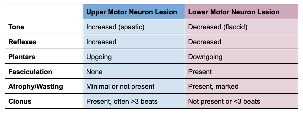
    Physical Exam Findings: Upper vs Lower Motor Neuron Lesions

    Physical Exam Findings: Upper vs Lower Motor Neuron Lesions

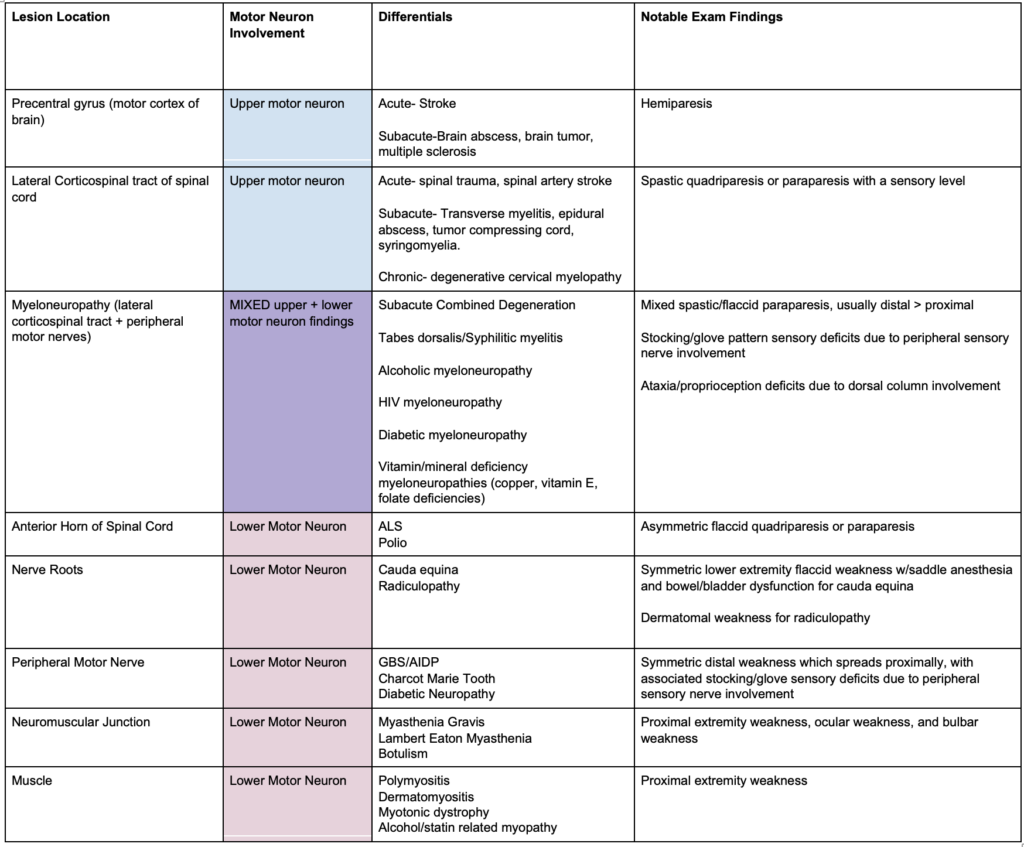
    Localize the Lesion- Differential Diagnoses for Extremity Weakness

    Localize the Lesion- Differential Diagnoses for Extremity Weakness

    Localize the Lesion- Differential Diagnoses for Extremity Weakness

    Localize the Lesion- Differential Diagnoses for Extremity Weakness

    MRI Findings and Subacute Combined Degeneration

    • The MRI displayed symmetric high signal intensity in the dorsal columns, a diagnostic feature identified as the inverted V sign or inverted rabbit ear sign.
    • Significance of the Inverted V Sign: This MRI sign is pathognomonic for subacute combined degeneration, indicating it is a distinct marker for this condition.
    • T2 Weighted Axial Images: The inverted V sign is observed in T2 weighted axial MRI images, which are used to evaluate the presence and extent of demyelination within the spinal cord.
    • Interpretation of Hyperintense Signals: Hyperintense signals on T2 weighted images generally indicate demyelination, where the protective myelin sheath around nerve fibers is damaged or destroyed.
    • Anatomical Location: The dorsal columns, located anatomically dorsal (toward the back) within the spinal cord, will appear toward the bottom of the screen in an axial (cross-sectional) view on the MRI.
    • Demyelination Appearance: Demyelination in the dorsal columns, typically situated in the thoracic spine, manifests as an upside-down V shape on the MRI, correlating with the described inverted V or rabbit ear sign.

    Pathophysiology of SCD due to Nitrous Oxide

    • Nitrous Oxide’s Effect on Vitamin B12: Nitrous oxide inactivates vitamin B12 by oxidizing a cobalt component within the molecule, rendering the vitamin functionally ineffective despite adequate consumption and absorption.
    • Impact on Methionine Synthase: The oxidation of vitamin B12 by N2O prevents it from activating methionine synthase, an enzyme critical for important biochemical processes.
    • Folate to Tetrahydrofolate Conversion: Inactive methionine synthase cannot convert folate into tetrahydrofolate, which is necessary for DNA synthesis. This disruption can lead to megaloblastic anemia, a condition associated with N2O-induced subacute combined degeneration.
    • Conversion of Homocysteine to Methionine: Methionine synthase is also responsible for converting homocysteine to methionine. Methionine is essential for the maintenance of myelin integrity, the protective sheath around nerve fibers.
    • Demyelination and Neurological Symptoms: The inability to maintain myelin integrity due to disrupted methionine production leads to the demyelination of dorsal columns and peripheral motor/sensory nerves, characteristic of N2O-SCD.
    • Normal B12 Levels with Functional Deficiency: Blood levels of vitamin B12 can appear normal in individuals affected by N2O exposure, as the issue lies in the vitamin’s inactivation rather than its absence, creating a functional deficiency.
    • Diagnosis of N2O-SCD: To diagnose N2O-induced SCD, healthcare providers need to check for elevated levels of methylmalonic acid and homocysteine. These substances are typically metabolized with the help of vitamin B12, and their elevated levels indicate a functional deficiency of B12 due to N2O exposure.

    Treatment and Management

    • Lack of Standardized Treatment: There is no universally accepted treatment protocol for N2O induced SCD, but common practices exist based on neurologist recommendations.
    • B12 Injection Protocol: A common approach involves administering vitamin B12 injections daily or every other day until there is noticeable improvement in symptoms. Once symptoms start to improve, the frequency of injections can be reduced to once a week.
    • Importance of Abstinence from N2O: For recovery to be possible, it is crucial that the patient completely abstains from using whippets (recreational N2O canisters). Continuing to use N2O can inactivate the administered vitamin B12, undermining the treatment efforts.
    • Recovery Process: Recovery from N2O induced SCD is typically slow and may not be complete. While remyelination and neurological function can gradually improve, the process is lengthy and may not fully return to baseline.
    • Recovery Statistics: Approximately 80% of individuals with N2O-SCD experience some improvement after a year of consistent B12 treatment. However, only between 10% and 20% of patients fully recover to their pre-condition baseline.
    • Risk Factors and Prevalence: The risk of developing SCD correlates with the frequency and quantity of N2O use. About 3.4% of individuals who use whippets will develop SCD, with the risk increasing to 8.5% among those who use more than 100 canisters per session. The case in point involved a patient using 20-40 canisters per session.
    • Increased Risk with Preexisting Conditions: Individuals who already have a vitamin B12 deficiency are at a greater risk of experiencing SCD symptoms, even with minimal use of whippets. This highlights the importance of understanding individual health conditions and potential vulnerabilities when assessing risk.

    Conclusion and Preventive Measures

    • Providers should be vigilant in screening for nitrous oxide use among patients presenting with unexplained neurological symptoms. The goal is to enhance early detection and treatment of N2O-induced SCD and to educate patients on the potential long-term health consequences of recreational nitrous oxide use.

    References

    • Neurology. Mumenthaler M, Mattle H, Taub E, ed. 4th Edition. Stuttgart: Thieme; 2003. doi:10.1055/b-005-148905
    • Zayia LC, Tadi P. Neuroanatomy, Motor Neuron. [Updated 2023 Jul 24]. In: StatPearls [Internet]. Treasure Island (FL): StatPearls Publishing; 2024 Jan-. Available from: https://www.ncbi.nlm.nih.gov/books/NBK554616/
    • Khin-Htun S, Tan H, Aung T. “Approach to a Patient With Weakness” 2021 Feb 15. Youtube. https://www.youtube.com/watch?v=3WQvtCuC4Fo&t=922s
    • Bhattacharyya S.Spinal Cord Disorders: Myelopathy, The American Journal of Medicine, Volume 131, Issue 11, 2018, Pages 1293-1297, ISSN 0002-9343,https://doi.org/10.1016/j.amjmed.2018.03.009.
    • ​​Garg RK, Malhotra HS, Kumar N. Approach to a case of myeloneuropathy. Ann Indian Acad Neurol. 2016 Apr-Jun;19(2):183-7. doi: 10.4103/0972-2327.182303. PMID: 27293327; PMCID: PMC4888679.
    • Lim PAC. Transverse Myelitis. Essentials of Physical Medicine and Rehabilitation. 2020:952–9. doi: 10.1016/B978-0-323-54947-9.00162-0. Epub 2019 Apr 17. PMCID: PMC7151963.
    • Jayarangaiah A, Lui F, Theetha Kariyanna P. Lambert-Eaton Myasthenic Syndrome. [Updated 2023 Oct 23]. In: StatPearls [Internet]. Treasure Island (FL): StatPearls Publishing; 2024 Jan-. Available from: https://www.ncbi.nlm.nih.gov/books/NBK507891/
    • Nguyen TP, Taylor RS. Guillain-Barre Syndrome. [Updated 2023 Feb 7]. In: StatPearls [Internet]. Treasure Island (FL): StatPearls Publishing; 2024 Jan-. Available from: https://www.ncbi.nlm.nih.gov/books/NBK532254/
    • Froese DS, Fowler B, Baumgartner MR. Vitamin B12 , folate, and the methionine remethylation cycle-biochemistry, pathways, and regulation. J Inherit Metab Dis. 2019 Jul;42(4):673-685. doi: 10.1002/jimd.12009. Epub 2019 Jan 28. PMID: 30693532.
    • Guo CJ, S. Kaufman B. Inhalational Anesthetics. In: Nelson LS, Howland M, Lewin NA, Smith SW, Goldfrank LR, Hoffman RS. eds. Goldfrank’s Toxicologic Emergencies, 11e. McGraw-Hill Education; 2019. Accessed February 27, 2024. https://accessemergencymedicine-mhmedical-com.ezproxy.med.nyu.edu/content.aspx?bookid=2569&sectionid=210274345
    • Lin JP, Gao SY, Lin CC. The Clinical Presentations of Nitrous Oxide Users in an Emergency Department. Toxics. 2022 Feb 26;10(3):112. doi: 10.3390/toxics10030112. PMID: 35324737; PMCID: PMC8950993.
    • Qudsiya Z, De Jesus O. Subacute Combined Degeneration of the Spinal Cord. [Updated 2023 Feb 12]. In: StatPearls [Internet]. Treasure Island (FL): StatPearls Publishing; 2023 Jan-. Available from: https://www.ncbi.nlm.nih.gov/books/NBK559316/
    • Hemmer B, Glocker FX, Schumacher M, et alSubacute combined degeneration: clinical, electrophysiological, and magnetic resonance imaging findingsJournal of Neurology, Neurosurgery & Psychiatry 1998;65:822-827.
    • Shah K, Murphy C. Nitrous Oxide Toxicity: Case Files of the Carolinas Medical Center Medical Toxicology Fellowship. J Med Toxicol. 2019 Oct;15(4):299-303. doi: 10.1007/s13181-019-00726-x. Epub 2019 Aug 6. PMID: 31388940; PMCID: PMC6825085.
    • Kalmoe MC, Janski AM, Zorumski CF, Nagele P, Palanca BJ, Conway CR. Ketamine and nitrous oxide: The evolution of NMDA receptor antagonists as antidepressant agents. J Neurol Sci. 2020 May 15;412:116778. doi: 10.1016/j.jns.2020.116778. Epub 2020 Mar 19. PMID: 32240970.
    • https://www.nytimes.com/2021/01/30/style/nitrous-oxide-whippets-tony-hsieh.html

    Read More

    Episode 193: Threatened Abortion Feb 01, 2024
    Show notes

    We review threatened abortion and the complexities in its care.

    Hosts:
    Stacey Frisch, MD
    Brian Gilberti, MD

    https://media.blubrry.com/coreem/content.blubrry.com/coreem/Threatened_Abortion.mp3 Download One Comment Tags: OBGYN

    Show Notes

    Background

    • Defined as vaginal bleeding during early pregnancy (before 20 weeks) with a closed cervical os, no passage of fetal tissue, and IUP on ultrasound
    • Occurs in 20-25% of all pregnancies.

    Initial Assessment and Management

    • Priority is to assess patient stability, establish good IV access, FAST may be helpful in identifying some ruptured ectopics early
    • Broad differential diagnosis is crucial to avoid mistaking conditions like ectopic pregnancy for other emergencies.
    • Importance of a detailed history and physical examination.

    Diagnostic Approach

    • Essential tests include HCG level, urinalysis, and possibly CBC + blood type/Rh status.
    • Rhogam’s use is well-supported in second and third trimester bleeding; however, data is less robust for first trimester bleeding in preventing sensitization
    • Importance of interpreting b-HCG with caution and understanding HCG discriminatory zones.
    • Use of ultrasound imaging, both bedside and formal, to assess the pregnancy’s status.

    Patient Counseling and Management

    • Open and honest communication about the prognosis of threatened abortion.
    • Addressing psychosocial aspects, including dispelling guilt and myths, and screening for intimate partner violence and mental health issues.
    • Recommendations against bedrest and certain activities
    • Lack of evidence supporting restrictions on sexual activity.
    • Standard pregnancy guidelines: avoiding smoking, alcohol, drug use, and starting prenatal vitamins.

    Follow-up and Precautions

    • Adopting a wait-and-see approach for stable patients, with scheduled follow-ups for ultrasounds and beta-HCG tests.
    • Educating patients on critical warning signs that require immediate medical attention.
    • Emphasizing the importance of returning to the hospital if experiencing significant bleeding or other severe symptoms.

    Take Home Points

    1. Threatened Abortion is defined as Experiencing abdominal pain and/or vaginal bleeding during early pregnancy (before 20 weeks), characterized by a closed cervical os and no expulsion of fetal tissue. In these cases, it is important to assess patient stability promptly.
    2. Keep your differential broad in these cases. The evaluation will in most cases involve a combination of labs and ultrasound imaging.
    3. Understand that the Rhogam certainly has a role in second and third trimester vaginal bleeding in the Rh-negative patient, and that there is a dearth of good data on its role in the first trimester – it will ultimately be a decision that is made by you, OBGYN, and the patient.
    4. Approach the interpretation of HCG levels with caution and remember that ectopic pregnancies might not adhere to conventional HCG levels.
    5. Established follow up and discharge instructions are crucial. Manage stable patients with a watchful waiting approach, scheduling subsequent visits for continuous ultrasounds and HCG testing. Clearly outline the importance of immediate medical attention for symptoms such as intense bleeding, significant abdominal pain, fever, or feelings of insecurity at home.
    6. Finally, we play an important role wherein we must ensure that the patient is medically stable and psychosocially safe. Here, compassionate communication is crucial when discussing what the diagnosis might entail, alleviate any feelings of blame or shame, and remain vigilant for signs of intimate partner violence or mental health issues. As emergency medicine physicians, it’s crucial for us to approach these cases with a comprehensive mindset.

    Read More

    Episode 192: Syncope in Children Jan 03, 2024
    Show notes

    We review a general approach to syncope in children.

    Hosts:
    Brian Gilberti, MD
    Ellen Duncan, MD

    https://media.blubrry.com/coreem/content.blubrry.com/coreem/Syncope_in_Children.mp3 Download Leave a Comment Tags: Cardiology, Pediatrics

    Show Notes

    • Initial Evaluation and Management:
      • Similar initial workup for children and adults: checking glucose levels for hypoglycemia and conducting an EKG.
      • The history and physical exam are crucial.
    • Dextrose Administration in Children:
      • Explanation of the ‘rule of 50s’ for determining the appropriate dextrose solution and dosage for children.
    • ECG Analysis:
      • Importance of ECG in diagnosing dysrhythmias like long QT syndrome, Brugada syndrome, catecholamine polymorphic V tach, ARVD, ALCAPA, and Wolff-Parkinson-White syndrome.
      • Younger children’s dependency on heart rate for cardiac output and the risk of arrhythmias in kids with congenital heart disease.
    Condition Characteristic ECG Findings Congenital/Acquired
    Long QT Syndrome (LQTS) Prolonged QT interval Congenital/Acquired
    Wolff-Parkinson-White Syndrome (WPW) Short PR interval, Delta wave Congenital
    Brugada Syndrome ST elevation in V1-V3, Right bundle branch block Congenital
    Atrioventricular Block (AV Block) PR interval prolongation (1st degree), Missing QRS complexes (2nd & 3rd degree) Congenital/Acquired
    Supraventricular Tachycardia (SVT) Narrow QRS complexes, Absence of P waves, Tachycardia Congenital/Acquired
    Ventricular Tachycardia Wide QRS complexes, Tachycardia Congenital/Acquired
    Arrhythmogenic Right Ventricular Dysplasia (ARVD/C) Epsilon waves, V1-V3 T wave inversions, Right bundle branch block Congenital
    Hypertrophic Cardiomyopathy (HCM) Left ventricular hypertrophy, Deep Q waves Congenital
    Pulmonary Hypertension Right ventricular hypertrophy, Right axis deviation Acquired
    Athlete’s Heart Sinus bradycardia, Voltage criteria for left ventricular hypertrophy Acquired
    Catecholaminergic Polymorphic VT (CPVT) Bidirectional or polymorphic VT, typically normal at rest Congenital
    Anomalous Origin of Left Coronary Artery from Pulmonary Artery (ALCAPA) May be normal, signs of ischemia or infarction in severe cases Congenital
    • History Taking:
      • Key aspects include asking about syncope with exertion, syncope after being startled, and syncope after pain or emotional stress.
      • Prolonged loss of consciousness may indicate seizures, and emotional stress and pain can trigger breath-holding spells.
    • Breath-Holding Spells:
      • Clarification of misconceptions about breath-holding spells, discussing their causes and characteristics, like cyanotic and pallid types.
      • Association with iron deficiency and the fact that most children outgrow these spells by age 8.
    • Physical Examination and History:
      • A cardiac exam is vital, with specific signs to look for, like murmurs in hypertrophic cardiomyopathy.
      • History can help identify the etiology of syncope, such as vasovagal responses or orthostatic hypotension.
    • Vasovagal Syncope:
      • Common in kids, especially teenagers, typically presenting with a prodrome of lightheadedness, diaphoresis, and pallor.
      • Normal glucose and EKG are expected in these cases.
    • Additional Lab Tests:
      • Pregnancy tests in reproductive-age women, and checking for less common causes like pulmonary embolism, subarachnoid hemorrhage, and toxic exposures.

    Take Home Points:

    • Immediate assessments for syncope in children should include a FS to evaluate for hypoglycemia and an ECG to evaluate any cardiac rhythm or conduction abnormalities.
    • Apply the “Rule of 50s” for hypoglycemic patients to suggest which fluids should be used.
    • Refer to our table for ECG findings to look out for when reviewing ECG tracings for these patients.
    • Pay particular attention to clues in the history that would suggested HCOM or seizures.
    • Breath-holding spells usually resolve by eight
    • HCOM murmurs will increase with Valsalva maneuver
    • Always keep your differential broad when approaching these patients given the heterogeneity of potential pathology that could lead to this chief complaint

    Read More

    Episode 191: Rapid Atrial Fibrillation Dec 01, 2023
    Show notes

    We go over the treatment of rapid atrial fibrillation (afib with RVR).

    Hosts:
    Brian Gilberti, MD
    Jonathan Kobles, MD

    https://media.blubrry.com/coreem/content.blubrry.com/coreem/Rapid_Atrial_Fibrillation.mp3 Download One Comment Tags: Cardiology

    Show Notes

    Understanding AF with RVR Categories

    • General AF with RVR: Definition and basic understanding.
    • Rapid AF with Pre-excitation: Characteristics and complications.
    • Chronic AF in Critical Illness: Identification and special considerations.

    Stability Assessment in AF with RVR

    • ACLS Protocols: Distinction between unstable and stable patients.
      • Unstable Patients: Immediate need for synchronized cardioversion, standard dose at 200 J for adults.
      • Stable Patients: Rate vs. rhythm control strategies, consideration of underlying etiology.
    • Limitations in Chronic AF: Challenges in patients with AF secondary to critical illness.

    ACLS Guidelines and ECG Findings

    • Tachycardia with a Pulse Approach: Initial assessment guidelines.
    • ECG Interpretation:
      • Irregularly Irregular Rhythm: Absence of discernible P waves.
      • Ventricular Rate: Typically over 100 bpm.
      • QRS Complexes: Usually narrow, alterations in the presence of bundle branch block or ventricular rate-related aberrancy.
    • Identifying Pre-Excitation Syndromes: Signs of shortened PR interval and slurred QRS, indication of Wolff-Parkinson-White Syndrome.

    AF with Pre-Excitation (WPW Syndrome)

    • Risk Assessment: Dangers of using AV nodal blockers (BB/CCB, digoxin, adenosine).
    • Alternative Management: Utilization of procainamide for stable patients, synchronized electrical cardioversion for unstable patients.

    Treatment Approaches for AF Types

    • General Rapid AF:
      • First Line Agents: Metoprolol vs. Diltiazem.
      • Metoprolol Considerations: Dosing (5 mg every 10-15 minutes, max 15 mg), benefits in CAD and HF, limitations in asthma/COPD patients.
      • Diltiazem Advantages: Faster action, suitability in asthma/COPD, typical dosing (0.25 mg/kg initial, followed by 0.35 mg/kg if needed).
    • Critically Ill Patients: Tailoring treatment to underlying pathology, avoiding typical AF pharmacologic treatments.

    Systematic Evaluation of Tachycardia Causes (TACHIES Mnemonic)

    • Thyrotoxicosis, Alcohol withdrawal, Cardiac issues, Hemorrhage, Intervals (WPW), Embolus, Sepsis.
    • Application of the mnemonic for a comprehensive approach to differential diagnosis.

    Ultrasound in Diagnostic Assessment

    • Application in Undiagnosed Tachycardia: Identifying EF, pericardial effusion, valvular pathology, and signs of pulmonary embolism.
    • Fluid Status Evaluation: Use of ultrasound for assessing b-lines in lung scans.

    Management of Chronic AF with HD Instability

    • Assessment of Hemodynamic Impact: Effects of extreme tachycardia on cardiac output, preload and afterload considerations.
    • Chronic vs. Paroxysmal AF: Differentiation in clinical presentation and treatment response.

    Approaches in Complex AF Cases

    • Addressing RVR of Unclear Etiology: Targeted therapies based on suspected underlying causes.
    • Medication Strategies:
      • Amiodarone: Bolus and drip approach, slow AV nodal without significant impact on contractility.
      • Esmolol: Titration for heart rate control, short-acting nature allowing for rapid cessation if adverse effects are observed.

    Comprehensive Patient Disposition

    • Considerations: Hemodynamic stability, underlying cause, comorbidities, outpatient follow-up feasibility.
    • Decision-Making Process: Balancing acute management with long-term treatment strategies.

    Take Home Points

    • Differentiation in AF with RVR Types: It’s essential to distinguish between primary AF with RVR, chronic AF with RVR related to other health issues, and new-onset AF (NOAF) with RVR in critically ill patients, as each type necessitates a unique approach to treatment.
    • ACLS Guidelines for AF with RVR: The ACLS guidelines provide a treatment framework, particularly recommending immediate synchronized cardioversion for unstable patients. However, these guidelines may have limited effectiveness for chronic AF with RVR patients suffering from underlying critical illnesses.
    • ECG Diagnosis in AF: Identifying AF on an ECG is crucial, with key indicators being an irregular rhythm without clear P waves and a ventricular rate exceeding 100 bpm. Accurate ECG interpretation guides effective treatment planning.
    • Special Cases like WPW Syndrome: WPW syndrome and similar conditions require careful treatment consideration, as standard AF treatments can worsen these conditions. Alternatives like procainamide or amiodarone are often more appropriate.
    • Patient-Centered Management of AF with RVR: Management should account for the patient’s overall health, underlying conditions, the chronicity of AF, and other comorbidities. Drugs like metoprolol and diltiazem offer benefits and risks, demanding personalized treatment plans.
    • Pathophysiology in Critical AF Patients: Understanding the underlying pathophysiology in critically ill patients is vital. Tachycardia in these cases might be compensatory, necessitating an investigation into causes like myocarditis, dehydration, or GI bleeding.
    • Systematic Evaluation with TACHIES Mnemonic: The mnemonic TACHIES (Thyrotoxicosis, Alcohol withdrawal, Cardiac issues, Hemorrhage, Intervals [WPW], Embolus, Sepsis) aids in systematically assessing and addressing emergent tachycardia causes in critically ill patients.

    Read More

    Episode 190: Electrical Storm Nov 01, 2023
    Show notes

    We discuss Electrical Storm (VT storm) and how to care for the very irritable heart.

    Hosts:
    Brian Gilberti, MD
    Reed Colling, MD

    https://media.blubrry.com/coreem/content.blubrry.com/coreem/Electrical_Storm.mp3 Download Leave a Comment Tags: Cardiology

    Show Notes

    Background/Overview of VT:

    • Definition: What makes it a storm
      • Three or more sustained episodes of VF, VT, or appropriate ICD shocks in a 24-hour period
    • Pathophysiology: Understanding the origin and mechanism
      • Sympathetic drive/adrenergic surge
      • Underlying pathology: Sodium channelopathies, infiltrative disease like cardiac sarcoidosis, etc.
    • RF’s / trigger / population (reversible cause in ~25% of patients)
      • MI
      • Electrolyte Derangements (emphasis on potassium and magnesium)
      • New/worsening heart failure
      • Catecholamine Surge
      • Drugs (stimulants, cocaine, amphetamines, etc)
      • QT Prolongation
      • Thyrotoxicosis

    Clinical Presentation:

    • Symptoms of VT: spectrum of symptoms – from palpitations to syncope to cardiac arrest
    • Differentiating VT from other potential ER presentations.

    Diagnostics in ER:

    • Electrocardiogram (ECG): Recognizing VT patterns.
      • Monomorphic vs polymorphic (Torsades) may change management
      • Wide QRS
      • Fusion best
      • Capture beats
      • Concordance
      • AV-dissociation
    • Lab tests: Potassium, magnesium, troponins, TFTs, etc.

    Acute Management in the ER:

    • Hemodynamically stable vs. unstable V
      • Unstable = cardioversion
      • Sedation
        • Catecholamine surge should be considered
        • No ideal agent
        • Etomidate or propofol can be considered
        • Ketamine may worsen irritability
    • Pharmacological treatments:
      • Amiodarone
        • Class III antiarrhythmic
        • Most studied in VT storm
        • First line
      • Beta Blockers
        • Propranolol
        • B1 and B2 activity
    • Non-pharmacological approaches:
      • Immediate synchronized cardioversion
      • IABP / ECMO considered for HD unstable patient
      • Cath lab if ischemic etiology suspected
      • Stellate Ganglion Block

    Take Home Points

    • Definition: VT Storm is commonly defined as three or more sustained episodes of ventricular fibrillation, ventricular tachycardia, or appropriate ICD shocks within a 24-hour period.
    • Varied Presentation: Patients may experience a range of symptoms from palpitations to severe hemodynamic instability.
    • ECG and Diagnosis: Initial ECG may not show VT; continuous cardiac monitoring or device interrogation may be required for diagnosis.
    • VT Identification: Look for wide QRS, rate over 100, fusion beats, capture beats, and AV dissociation to identify VT.
    • Management in Hemodynamic Instability: Cardiovert if the patient shows signs of hemodynamic instability.
    • Sedation Considerations: Be cautious with sedation, especially with ketamine, as it may worsen cardiac irritability in these already adrenergic state patients.
    • Medication Choices: Typically, amiodarone and propranolol are used to manage VT Storm.
    • Cardiology Involvement: Involve cardiology early on, as treatment may extend beyond medications.

    Read More

    Episode 189: Hyperkalemia 2.0 Oct 01, 2023
    Show notes

    We revisit the topic of Hyperkelamia to update our prior episode from 2015 (pre-Lokelma)

    Hosts:
    Brian Gilberti, MD
    Jonathan Kobles, MD

    https://media.blubrry.com/coreem/content.blubrry.com/coreem/Hyperkalemia.mp3 Download 2 Comments Tags: Renal Colic

    Show Notes

    Introduction

    1. Background
      • Physiology:
        • Normal range and the significance of deviations (>5.5 mEq/L)
      • Epidemiology:
        • Prevalence of hyperkalemia in the ER
        • ESRD missed HD → ECG, monitor

    Causes / Risk Factors

      • Causes
        • Kidney Dysfunction, Medications, Cellular Destruction, Endocrine Causes, Pseudohyperkalemia
      • High-Risk Medications:
        • Antibiotics: Bactrim, antifungals
        • Calcineurin inhibitors
        • Beta-blockers
        • ACE/ARB
        • K+ Sparing diuretics
        • NSAIDs
        • Digoxin
        • SUX – high risks in neuromuscular disease
      • Lab errors, hemolysis in samples
        • VBG vs Chem accuracy
        • When to repeat a hemolyzed sample
        • 2023 study: Of the 145 children with hemolyzed hyperkalemia, 142 (97.9%) had a normal repeat potassium level. Three children (2.1%) had true hyperkalemia: one had known chronic renal failure and was referred to the ED due to concern for electrolyte abnormalities; the other 2 patients had diabetic ketoacidosis (DKA).

    Clinical Presentation / eval

    • Symptomatic vs. Asymptomatic:
      • “First symptom of hyperkalemia is death”
      • If severe, ascending muscle weakness → paralysis
        • Point at which patients experience symptoms depends on chronicity
          • >7 mEq/L if chronic and can be lower if acute
      • Hyperkalemia can be a cause of non-specific GI symptoms
    • EKG Changes:
      • ECG findings may be the first marker the ER doc gets that something is wrong
      • Typical changes:
        • Peaked T-waves, shortened QT
        • Lengthening of PR interval and QRS duration
        • Bradycardia / Junctional rhythm
          • Hyperkalemia can produce bradycardia without other ECG findings
        • Ones associated with VT/VF/code, death in one study: QRS widening (RR = 4.74), Junctional Rhythm (RR = 7.46), HR <50 (RR = 12.29) while no adverse outcomes with just peaked T waves or PR prolongation (Durfey, 2017)
      • Don’t be fooled by a normal ECG, may be normal, but it’s also on case report level to have K > 9 and a normal ECG
        • Series of 127 patient (K 6-9.3), no serious arrhythmia noted, only 46% had ECG changes, (Acker, 1998)
      • ECG changes are not linear, there is no exact association between K+ levels and ECG changes
      • ECG changes may be hidden and subtle in patients with underlying inter-ventricular conduction delay (BBBs)
        • Be suspicious of the patient with LBBB > 160 ms or RBBB > 140 ms
      • BRASH Syndrome
        • Synergism between hyperkalemia, renal failure/injury and AV nodal blocking agents -> may produce ECG changes out of proportion to serum potassium levels.
    • Labs
      • Chem, VBG, +/- CK if you think muscle breakdown is at play (Tintinalli talks about looking at urine K, but this is not most people’s practice)
      • Consider evaluation for adrenal insufficiency
      • Waiting for labs may not be an option
        • Renal dysfunction + consistent ECG findings → prompt treatment before chem results
        • Realistically 2 hours to get back chemistry in most settings ≈ eternity

    Management in the ER

    • Discontinue/hold any nephrotoxins or medications in suspected medication-induced hyperkalemia
    • A. Acute Management Strategies:
      • Cardiac protection with calcium
        • 1g over 5-10 mins
          • Lasts 30-60 mins, may have to redose
          • Dose considerations if on digoxin
          • AEs: Calciphylaxis and hypercalcemia
            • Fast pushes can result in hypotension, arrhythmia
        • Calcium chloride vs calcium gluconate
        • Caution in patients taking Digoxin
      • IVF choice – NS vs LR
        • Caution/Avoid fluid in patients with ESRD/CHF or signs of VOL
      • Shifting potassium:
        • insulin/glucose
          • 5 units vs 10 units
            • 5 similar effect, less hypoglycemic episodes (LaRue 2017)
            • If doing 10 units, start D10W at 50-75 cc/h after amp of d50 but be mindful that anuric patient who missed HD may not have much room for volume
          • Decrease but about 0.5-1.2 mEq/L
          • Effect starts 10-20 mins after administration and can last 4-6 hours
        • Albuterol
          • 10-20 mg over 10 mins (NB: higher dose than for asthma)
          • Peak effect at 90 mins
          • Decreases by 0.5 – 1.0 mEq/L alone
            • With insulin, ~1.2 mEq/L, additive effect
        • Bicarbonate
          • Controversy. Useless in hyperkalemic, nonacidotic patient. Useful as drip but takes hours to work, again, volume in anuric patient an issue
            • May be most useful in patients with renal failure and hyperkalemia 2/2 volume loss
          • Hypertonic Bicarb is ineffective – More potassium is pulled out of cells due to osmotic shift.
      • Removal:
        • Lokelma (Sodium Zirconium cyclosilicate)
          • Luckily residents have never had to use Kayexalate
          • Can start working in 1-2 hours of administration
          • 0.37 mEq/L reduction at 4 hours after 10 g
          • Not a magic bullet in patients who need dialysis
        • Diuretics
          • No studies that demonstrate effectiveness in this ED setting
            • May be effective in patients with normal renal function
          • If patient not anuric, may be worth using, can give 40 mg, but again, should not be the only attempted method of removing K
          • Nephron BOMB
            • Loop Diuretic (160-250 mg IV Lasix or 4-5 mg IV Bymex)
            • Thiazide (500-1000 mg IV chlorothiazide or 5-10 mg metolazone)
            • +/- Acetazolamide
            • +/- Fludrocortisone
              • May help stimulate the kidneys to secrete potassium
              • Primarily helpful in patients with mineralocorticoid deficiencies
        • Dialysis
          • Involve renal early because it takes a while to call in an HD nurse sometimes
          • If no access and emergent HD is required → HD catheter placement
      • Strategies for suspected Brash syndrome
        • Epinephrine/Levo (if hypotensive/bradycardic)
        • Calcium gtt
    • Disposition/wrap up
      • Many factors at play here – patient preference, access, degree of hyperkalmia, identifiable / corrected cause

    Take Home points

      • Hyperkelamia causes can be put into three categories, pseudohyperkalemia, due to redistribution, and due to total body increase in potassium. Check out the show notes for a more complete list
      • Hyperkalemia can be difficult to pick up on before the labs come back because it can lurk without symptoms or even ECG changes
      • If a patient does have ECG changes, they may not follow that linear pattern that is traditionally taught and ECGs can be poorly sensitive. Now, if you do see changes, the ones that are more commonly associated with adverse events are QRS widening, junctional rhythm, and bradycardia
      • Treatment is a numbers game, calcium for cardiac stabilization can last just 30-60 minutes, insulin will be the fastest way to shift potassium back into cells, but be mindful that 10 units is associated with increased episodes of hypoglycemia whereas 5 units may have the same effect in reducing potassium. And albuterol is at a much higher dose than what is given for asthma
      • Lokelma is now a pillar of treatment for removal of potassium.
      • Diuretics with the goal of kiuresis may have a role in the oliguric patient, and increased doses along with other agents may buy time in patients with severe hyperK when HD is not readily available
      • Involve renal early if you think that the patient will require HD

    Read More

    Episode 188: Vasopressors Sep 01, 2023
    Show notes

    We go over the essential and complex topic of vasopressors in the ED.

    Hosts:
    Brian Gilberti, MD
    Catherine Jamin, MD

    https://media.blubrry.com/coreem/content.blubrry.com/coreem/Vasopressors.mp3 Download Leave a Comment Tags: Critical Care

    Show Notes

    Introduction

    • Host: Brian Gilberti, MD
    • Guest: Catherine Jamin, MD
      • Associate professor of Emergency Medicine at NYU Langone Health
      • Vice Chair of Operations
      • Triple-boarded in Emergency Medicine, Internal Medicine, and Critical Care Medicine
    • Topic: Vasopressors: Essential agents for supporting critically ill patients in the ED

    What Are Vasopressors and When to Use Them

    • Two primary mechanisms to increase blood pressure:
      1. Increasing systemic vascular resistance via vasoconstriction
      2. Increasing cardiac output via augmenting inotropy and chronotropy
    • Indicators for vasopressor use:
      • MAP <65, systolic BP <90, or significant drop from baseline BP
      • Signs of organ dysfunction like altered mental status, decreased urine output, elevated lactate
      • Fluid resuscitation either ineffective or contraindicated (e.g., in CHF patients)

    Commonly Used Vasopressors in the ED

    • Norepinephrine
    • Epinephrine
    • Vasopressin
    • Phenylephrine

    Norepinephrine

    • Mechanism: Stimulates alpha-1 (vasoconstriction) and beta-1 receptors (increases inotropy & chronotropy)
    • Starting Dose: 10 mcg/min, titrate to MAP >65
    • Max Dose: No strict limit but usually add a 2nd pressor at 15-20 mcg/min
    • Situational Preference: First-line for most cases of shock (septic, undifferentiated, hypovolemic, cardiogenic)
    • Pros: Can be infused peripherally via large bore IV

    Vasopressin

    • Mechanism: Activates V1a receptors causing vasoconstriction
    • Dose: Fixed, non-titratable dose of 0.04 units/min
    • Situational Preference: Second-line in septic shock
    • Concerns: Potential for peripheral ischemia

    Phenylephrine

    • Mechanism: Stimulates alpha-1 receptors causing vasoconstriction
    • Starting Dose: 100 mcg/min, titrate to MAP >65
    • Situational Preference: High cardiac output states, tachyarrhythmias, peri-intubation
    • Concerns: Increases afterload, can worsen low cardiac output states

    Epinephrine

    • Mechanism: Stimulates alpha-1, beta-1 and beta-2 receptors
    • Starting Dose: 5-10 mcg/min, titrate to MAP >65
    • Situational Preference: Anaphylactic shock, septic cardiomyopathy
    • Limitations: Can induce tachycardia, may elevate lactate levels

    Escalation Strategy in Refractory Shock

    • Norepinephrine -> Vasopressin (with stress dose steroids) -> Epinephrine
    • Consider POCUS, lactate, central venous saturation, and acid-base status

    Peripheral Pressors

    • Can safely be administered peripherally via large bore IVs in proximal upper extremity
    • Sites: Cephalic or basilic veins
    • Adverse Events: Low at 1.8% based on meta-analysis
    • Actions in case of extravasation: Phentolamine injection, nitroglycerin paste

    Push-Dose Pressors

    • Primarily Phenylephrine (peri-intubation, during procedures)
    • Also Epinephrine for peri-code situations
    • Doses: Epi – 5-20 mcg every 2-5 min

    Take-Home Points

    • Most used medications are going to be norepinephrine, vasopressin, phenylephrine, and epinephrine.
    • Consider these medications if there are signs of end-organ dysfunction, there is a considerable delta in baseline BP, systolic is less than 90 and/or MAP is less than 65
    • Norepinephrine is a good pressor for a lot of the situations that we encounter in the emergency department, such as septic shock, undifferentiated shock and hypovolemic shock.
    • Vasopressin is commonly the second we reach for in most of these scenarios
    • Epinephrine will be first for anaphylactic shock and may be the third agent in septic shock
    • Think about phenylephrine in high-output states (patients with tachydysrhythmias), or with AS, though be cautious in patient with low cardiac output
    • The benefits outweigh risks for peripheral pressors in situations where you promptly have to increase blood pressure while you work on central access
    • Push-dose pressures can help you in a peritinbatuion or pericode situation because it is going to be one of the fastest ways we can boost BP while we work on other measures to stabilize the patient

    Additional References

    • Importance of RUSH (Rapid Ultrasound in SHock) exam for diagnosis and treatment planning: https://emcrit.org/rush-exam/

    Read More

    Previous 1 2 3 4 5 6 24 Next

    Related Podcasts

    The Joe Rogan Experience

    1

    The Joe Rogan Experience Comedy
    More Perfect

    2

    More Perfect Government
    Casefile True Crime

    3

    Casefile True Crime Games & Hobbies
    Reply All

    4

    Reply All Games & Hobbies
    You Must Remember This

    5

    You Must Remember This Games & Hobbies
    Real Time with Bill Maher

    6

    Real Time with Bill Maher Games & Hobbies
    footer-logo

    Contact Us

    Toll Free: 844-670-7747

    Links

    • Home
    • Top Charts
    • Networks
    • Apps
    • Independents Podcasts
    • Podcast Advertising
    • Podcast News
    • Contact Us
    • About Us
    • Analytics & Insights

    Stay Connected

      Privacy, Terms of Use & Our Code of Ethics Protecting Content Creators Copyrights