TopPodcast.com
Menu
  • Home
  • Top Charts
  • Top Networks
  • Top Apps
  • Top Independents
  • Top Podfluencers
  • Top Picks
    • Top Business Podcasts
    • Top True Crime Podcasts
    • Top Finance Podcasts
    • Top Comedy Podcasts
    • Top Music Podcasts
    • Top Womens Podcasts
    • Top Kids Podcasts
    • Top Sports Podcasts
    • Top News Podcasts
    • Top Tech Podcasts
    • Top Crypto Podcasts
    • Top Entrepreneurial Podcasts
    • Top Fantasy Sports Podcasts
    • Top Political Podcasts
    • Top Science Podcasts
    • Top Self Help Podcasts
    • Top Sports Betting Podcasts
    • Top Stocks Podcasts
  • Podcast News
  • About Us
  • Podcast Advertising
  • Contact
Not in our directory?
Add Show Here
Podcast Equipment
Center

toppodcastlogoOur TOPPODCAST Picks

  • Comedy
  • Crypto
  • Sports
  • News
  • Politics
  • True Crime
  • Business
  • Finance

Follow Us

toppodcastlogoStay Connected

    View Top 200 Chart
    Back to Rankings Page
    Government & Organizations

    Core EM – Emergency Medicine Podcast

    Core EM Emergency Medicine Podcast

    Advertise

    Copyright: © Core EM

    • Apple Podcasts
    • Google Play
    • Spotify

    Latest Episodes:
    Episode 177.0 – Hemoptysis Feb 17, 2020
    Show notes

    An overview and management tips of hemoptysis in the ED.

    Hosts:
    Brian Gilberti, MD
    Audrey Bree Tse, MD

    https://media.blubrry.com/coreem/content.blubrry.com/coreem/Hemoptysis.mp3 Download One Comment Tags: Critical Care, Pulmonary

    Show Notes

    OVERVIEW:

    • Definition:
      • expectoration/ coughing of blood originating from tracheobronchial tree
    • Sources:
      • Bronchial arteries (90%): under systemic circulatory pressure to supply supporting structures of the lung → heavier bleeding
      • Pulmonary arteries (5%): under low pressure to supply alveoli → milder bleeding
      • Nonbronchial arteries (5%): intercostal arteries, coronary arteries, thoracic/ upper/ inferior phrenic arteries
    • Quantification:
      • Mild: <20mL/ 24h
      • Massive defined anywhere from >300mL-1L/ 24hr
      • Mortality: 38% for massive (>500mL/ 24hr) vs 4.5% for nonmassive
    • Etiology (in adults):
      • Infectious (most common):
        • Bronchitis
        • PNA (necrotizing, lung abscess)
        • TB
        • Viral
        • Fungal
        • Parasitic
      • Malignancy:
        • Primary lung cancer vs metastatic disease
      • Pulmonary:
        • Bronchiectasis
        • COPD
        • PE/ infarction
        • Bronchopleural fistula
        • Sarcoidosis
      • Cardiac:
        • Mitral stenosis
        • Tricuspid endocarditis
        • CHF
      • Rheumatological:
        • Goodpasture Syndrome
        • SLE
        • Vasculitis (Wegener’s, HSP, Behcet)
        • Amyloidosis
      • Hematological:
        • Coagulopathy/ thrombocytopenia/ platelet dysfunction
        • DIC
      • Vascular:
        • Pulmonary HTN
        • AA
        • Pulmonary artery aneurysm
        • Aortobronchial fistula
        • Pulmonary angiodysplasia
      • Toxins:
        • Anticoagulation/ aspirin/ antiplatelets
        • Penicillamine, amiodarone
        • Crack lung
        • Organic solvents
      • Trauma:
        • Tracheobronchial rupture
        • Pulmonary contusion
      • Other:
        • bronchoscopy/ lung biopsy
        • Pulmonary artery or central venous catheterization
        • Foreign body aspiration
        • Pulmonary endometriosis (catamenial hemoptysis)
        • Idiopathic (up to 25% of cases)
      • Pseudohemoptysis:
        • Sinusitis
        • Epistaxis
        • Rhinorrhea
        • Pharyngitis
        • URI
        • Aspiration
        • GIB

    WORKUP:

    • HPI:
      • CP, SOB
      • B symptoms: fever, weight loss, chills, night sweats
      • Lymphadenopathy
      • Timeframe: acute vs chronic
      • Prior lung/ renal/ cardiac disease
      • Recreational drug/ cigarette/ chemical exposures
      • travel/ infectious exposure
      • Medications
      • Any other sites of bleeding
      • Precipitating factors
      • Description of blood clots
      • Patients are unable to accurately estimate degree of bleeding
    • PE:
      • Petechiae, edema, ecchymosis, ulcers, clubbing (chronic lung disease)
      • Cardiopulmonary
      • Sputum samples
    • Labs:
      • CBC w/ diff, BMP, LFTs, coags, T&S
      • ABG
      • UA
      • Infectious workup if suspected: cultures, grain stains
    • Imaging:
      • CXR: 20% will be normal. May see tumour, cavity, effusion, infiltrate, PTX. Early pulmonary hemorrhage may present as infiltrate
      • CT: only for stable patients! May see bronchiectasis, cavitary lesions, acinar nodules, tumours
      • CTA: bronchial arteries, aneurysms, PE
      • ECHO: identify valvular abnormalities, signs of PE, aortic aneurysm
    • Bronchoscopy:
      • Not often performed in ED, but therapeutic & diagnostic
      • Allows direct visualization of tumours, foreign bodies, granulomas, infiltration, as well as local therapy (vasoconstrictive agents, stent/ balloon tamponade, electrocautery, procoagulants)

    MANAGEMENT:

    • Goals:
      • Control airway
      • Protect healthy lung
      • Identify and treat underlying cause
      • Stabilize hemodynamics with volume resuscitation
    • Provider precautions (respiratory & contact)
    • ABCs, close monitoring
      • Early airway management: massive hemoptysis, respiratory compromise, hypoxia, risk factors (elderly, AMS, coagulopathic)
      • 2 x suction, preoxygenation, patient positioned upright, >8Fr ETT to facilitate suctioning/ bronch
      • If bleeding side can be identified, consider “selective intubation” into nonbleeding lung to minimize further aspiration of blood and to provide ventilation
      • Life threat = asphyxiation, not exsanguination. ~Only 150cc anatomic dead space in major airways
    • 2 x large bore IVs
    • MTP prn vs volume resuscitation
    • “Bad lung down” in lateral position: theoretical belief to minimize reflux of blood into normal lung
    • Correct coagulopathy
    • Consider nebulized TXA for nonmassive hemoptysis (500mg w/ NS per neb)
      • Double-blind, randomized controlled trial in 2018
      • Nebulized TXA (500mg TID) vs placebo (normal saline) in hemodynamically stable adult patients admitted with mild hemoptysis (<200 mL/ 24hr) and no respiratory instability
        • Additional exclusion criteria included those with renal failure, hepatic failure, or coagulopathy
      • Assessed mortality and hemoptysis recurrence rate at 30 days and 1 year
      • 25 patients randomized to receive TXA nebs, 22 randomized to receive normal saline nebs
      • Results:
        • Resolution of hemoptysis within 5 days of admission was significantly higher in TXA-treated patients than placebo patients (96% vs 50%; P < 0.0005)
        • Mean hospital length of stay was shorter for TXA group (5.7 +- 2.5 days vs 7.8 +- 4.6 days; P = 0.046)
        • Fewer patients in TXA group required invasive procedures to control bleeding vs placebo group (0% vs 18.2%; P = 0.041)
        • No side effects were noticed in either group
    • Antibiotics if infectious
    • Bronchoscopy: local therapy (vasoconstrictive agents, stent/ balloon tamponade, electrocautery, procoagulants)
      • Rigid bronch for unstable patients to evacuate clots vs fiberoptic bronch for stable patients
    • Bronchial artery embolization (call IR early!)
    • May require lobectomy or pneumonectomy (consult thoracic surgery)

    DISPOSITION:

    • Low threshold for higher level of care: only mild, hemodynamically stable hemoptysis on floor
    • Discharge: only if certain regarding etiology in healthy, hemodynamically stable patients with scant, resolved hemoptysis, no coagulopathy, and reassuring workup
      • Ensure patients have reliable follow up and avoid smoking. Strict return precautions!

    REFERENCES:

    • Kiraly A, Pang P, Cheema N. Hemoptysis. In: Schaider J, Barkin R, Hayden S, Wolfe R, Barkin A, Shayne P, Rosen P. Rosen and Barkin’s 5-Minute Emergency Medicine Consult. 5th Edition. Philadelphia, PA: Wolters Kluwer; 2015; 504-505.
    • Nickson, C. Haemoptysis. Life in the Fastlane. [litfl.com/haemoptysis/]. Updated April 9, 2019. Retrieved February 10, 2020.
    • Wand O, Guber E, Guber A, Schochet GE, Israeli-Shani L, Shitrit D. Inhaled Tranexamic Acid for Hemoptysis Treatment: A Randomized Controlled Trial. Chest. December 2018; 154(6): 1379-1384.
    • Young WF. Hemoptysis. In: Cline, David,eds. Tintinalli’s Emergency Medicine Manual. 7th Edition. New York : McGraw-Hill Medical; 2011; 473-476.

    Read More

    Episode 176.0 – Pneumonia Updates Jan 27, 2020
    Show notes

    We go over the recent updates in the workup and management of pneumonia.

    Hosts:
    Brian Gilberti, MD
    Audrey Tse, MD

    https://media.blubrry.com/coreem/content.blubrry.com/coreem/Pneumonia_Updates.mp3 Download Leave a Comment Tags: Infectious Diseases, Pulmonary

    Show Notes

    2007 Infectious Diseases Society of America/American Thoracic Society Criteria for Defining Severe Community-acquired Pneumonia

    Validated definition includes either one major criterion or three or more minor criteria

    • Minor criteria
      • Respiratory rate > 30 breaths/min PaO2/FIO2 ratio<250
        Multilobar infiltrates Confusion/disorientation
      • Uremia (blood urea nitrogen level > 20 mg/dl)
      • Leukopenia* (white blood cell count , 4,000 cells/ml)
      • Thrombocytopenia (platelet count , 100,000/ml)
      • Hypothermia (core temperature , 368 C) Hypotension requiring aggressive fluid
      • resuscitation
    • Major criteria
      • Septic shock with need for vasopressors
      • Respiratory failure requiring mechanical ventilation

    A special thanks to our Infectious Diseases Editor:

    Angelica Cifuentes Kottkamp, MD
    Infectious Diseases & Immunology
    NYU School of Medicine


    Read More

    Episode 175.0 – Posterior Circulation Stroke Jan 13, 2020
    Show notes

    Diagnosing and managing one of our critical diagnoses - posterior stroke.

    Hosts:

    Mukul Ramakrishnan, MD
    Audrey Bree Tse, MD

    https://media.blubrry.com/coreem/content.blubrry.com/coreem/final_posterior_stroke_podcast_post_edit.mp3 Download 2 Comments Tags: Neurology, Posterior Stroke

    Show Notes

    See Dr. Newman-Toker demonstrate the HINTS exam here

    Kattah JC, Talkad AV, Wang DZ, Hsieh YH, Newman-Toker DE. HINTS to diagnose stroke in the acute vestibular syndrome: three-step bedside oculomotor examination more sensitive than early MRI diffusion-weighted imaging. Stroke. 2009 Nov;40(11):3504-10


    Read More

    Episode 174.0 – Homelessness Dec 16, 2019
    Show notes

    We discuss one of the most complex problems we face – Homelessness

    Hosts:
    Kelly Doran, MD
    Audrey Tse, MD
    Brian Gilberti, MD

    https://media.blubrry.com/coreem/content.blubrry.com/coreem/Homelessness.mp3 Download One Comment Tags: Social Emergency Medicine

    Show Notes

    Special Thanks To:

    Dr. Kelly Doran, MD MHS

    Ronald O. Perelman Department of Emergency Medicine at NYU Langone Health, NYC Health + Hospitals/ Bellevue

    ___________________________

    References:

    Doran, K.M. Commentary: How Can Emergency Departments Help End Homelessness? A Challenge to Social Emergency Medicine. Ann Emerg Med. 2019;74:S41-S44.

    Doran, K.M., Raven, M.C. Homelessness and Emergency Medicine: Where Do We Go From Here? Acad Emerg Med. 2018;25:598-600.

    Salhi, B.A., et al. Homelessness and Emergency Medicine: A Review of the Literature. Acad Emerg Med. 2018;25:577-93.

    U.S. Department of Housing and Urban Development, Annual Homeless Assessment Report to Congress. Available at: https://www.hudexchange.info/resource/5783/2018-ahar-part-1-pit-estimates-of-homelessness-in-the-us/

    U.S. Interagency Council on Homelessness. Home, Together Federal Strategic Plan to Prevent and End Homelessness. https://www.usich.gov/resources/uploads/asset_library/Home-Together-Federal-Strategic-Plan-to-Prevent-and-End-Homelessness.pdf


    Read More

    Episode 173.0 – Blunt Neck Trauma Nov 25, 2019
    Show notes

    We go into one of the more complex injuries – blunt neck trauma.

    Hosts:
    Audrey Bree Tse, MD
    Brian Gilberti, MD

    https://media.blubrry.com/coreem/content.blubrry.com/coreem/Blunt_Neck_Injuries.mp3 Download One Comment Tags: Trauma

    Show Notes

    Overview

    • Blunt neck trauma comprises 5% of all neck trauma
    • Mortality due to loss of airway more so than hemorrhage

    Mechanism

    • MVCs with cervical hyperextension, flexion, rotation during rapid deceleration, direct impact
    • Strangulation: hanging, choking, clothesline injury (see section on strangulation in this chapter)
    • Direct blows: assault, sports, falls

    Initial Management/Primary Survey

    • Airway
      • Evaluate for airway distress (stridor, hoarseness, dysphonia, dyspnea) or impending airway compromise
      • Early aggressive airway control: low threshold for intubation if unconscious patient, evidence of airway compromise including voice change, dyspnea, neurological changes, or pulmonary edema
      • Assume a difficult airway
    • Breathing
      • Supplemental oxygen
      • Assess for bilateral breath sounds
      • Can use bedside US to evaluate for pneumothorax or hemothorax
    • Circulation
      • Assess for open wounds, bleeding, hemorrhage
      • IV access
    • Disability
      • Maintain C-spine immobilization
      • Calculate GCS
      • Look for seatbelt sign

    Secondary Survey

    • Evaluate for specific signs of vascular, laryngotracheal, pharyngoesophageal, and cervical spinal injuries with inspection, palpation, and auscultation
    • Perform extremely thorough exam to evaluate for any concomitant injuries (e.g. stab wounds, gunshot wounds, intoxications/ ingestions, etc.)

    Types of Injuries

    • Vascular injury
      • Overview
        • Carotid arteries (internal, external, common carotid) and vertebral arteries injured
        • Mortality rate ~60% for symptomatic blunt cerebral vascular injury
      • Mechanism
        • Hyperextension and lateral rotation of the neck, direct blunt force, strangulation, seat belt injuries, and chiropractic manipulation
        • Morbidity due to intimal dissections, thromboses, pseudoaneurysms, fistulas, and transections
      • Clinical Features
        • Most patients are asymptomatic and do not develop focal neurological deficits for days
        • if Horner’s syndrome, suspect disruption of thoracic sympathetic chain (wraps around carotid artery)
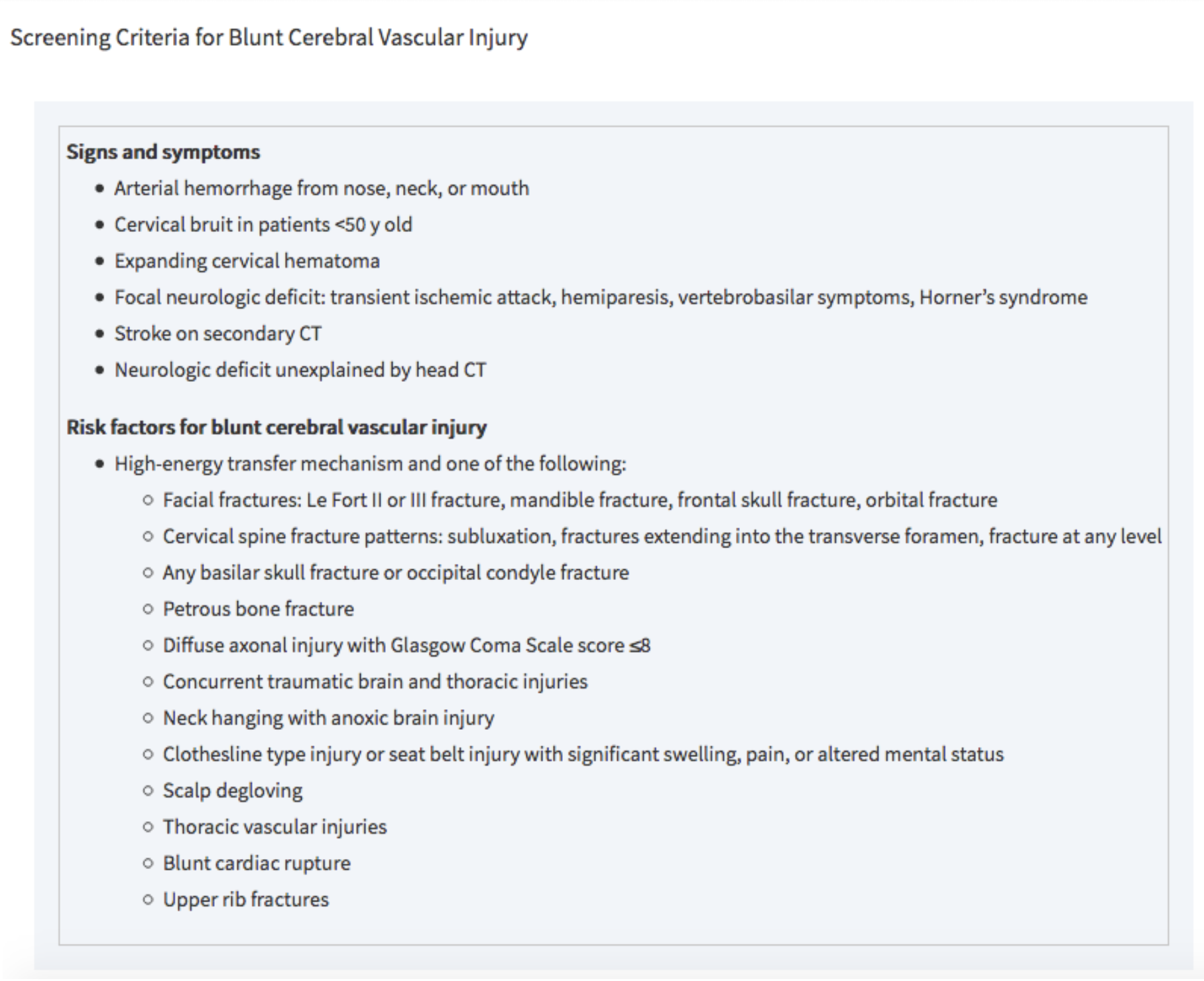
        • specific screening criteria are used to detect blunt cerebrovascular injury in asymptomatic patients (see below)

    Tintinalli 2016

    • Diagnostic Testing
      • Gold standard for blunt cerebral vascular injury = MDCTA (multidetector four-vessel CT angiography)
        • <80% sensitive but 97% specific
        • Also images aerodigestive tracts and C-spine (unlike angiography)
      • Followed by Digital Subtraction Angiography (DSA) for positive results or high suspicion
        • Angiography is invasive, expensive, resource-intensive, and carries a high contrast load
    • Management
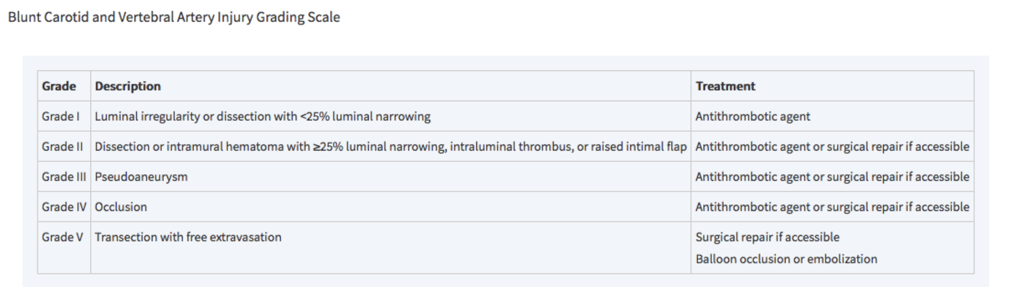
      • Antithrombotics vs. interventional repair based on BCVI grading system
      • Involve consultants early: trauma surgery, neurosurgery, vascular surgery, neurology
      • All patients with blunt cerebral vascular injury will require admission

    Tintinalli 2018

    • Pharyngoesophageal injury
        • Overview
          • Rare in blunt neck trauma
          • Includes hematomas and perforations of both pharynx and esophagus
        • Mechanism
          • Sudden acceleration or deceleration with hyperextension of the neck
          • Esophagus is thus forced against the spine
        • Clinical Features
          • Dysphagia, odynophagia, hematemesis, spitting up blood
          • Tenderness to palpation
          • SC emphysema
          • Neurological deficits (delayed presentation)
          • Infectious symptoms (delayed presentation)
        • Diagnostic Testing
          • Esophagography with water-soluble contrast (e.g. Gastrograffin)
          • If negative contrast esophagography, obtain flexible endoscopy (most sensitive)
            • Combination of contrast esophagography + esophagoscopy has sensitivity close to 100%
          • Swallow studies with water-soluble agent
          • MDCTA
          • Plain films of neck and chest
            • Findings such as pneumomediastinum, hydrothorax, or retropharyngeal air may suggest perforation but are not sensitive
        • Management
          • All pharyngoesophageal injuries receive IV antibiotics with anaerobic coverage
          • Parenteral/ enteral nutrition
          • NGT should only be placed under endoscopic guidance to avoid further injury
          • Medical management vs. surgical repair depending on extent of injury
            • Surgical repair for esophageal perforations or pharyngeal perforations >2cm
          • Involve consultants early: trauma surgery, vascular surgery, otolaryngology, gastroenterology
          • All patients with blunt cerebral vascular injury will require admission
    • Laryngotracheal injury
      • Overview
        • Occurs in >0.5% of blunt neck trauma
        • Includes hyoid fractures, thyroid/ cricoid cartilage damage, cricotracheal separation, vocal cord disruption, tracheal hematoma or transection
      • Mechanism
        • Assault, clothesline injuries, direct blunt force from MVCs compressing the larynx between a fixed object and the spine
      • Clinical Features
        • Patients are often asymptomatic at first and then develop airway edema and/or hematoma resulting in airway obstruction
        • Children are at higher risk for airway compromise due to less cartilage calcifications
      • Diagnostic Testing
        • Flexible fiberoptic laryngoscopy (FFL) to assess airway patency and extent of intraluminal injury
        • MDCTA
          • Obtain 1-mm cuts of larynx and perform multiplanar reconstructions
        • Consider POCUS to detect laryngotracheal separation
      • Plain films of neck and chest
      • Poor sensitivity for penetrating neck trauma injuries
      • Can show extraluminal air, fracture or disruption of cartilaginous (e.g. larynx) structures
      • Management
        • When securing airway, use an ETT that is one size smaller due to likelihood of airway edema
        • Conservative management (IV antibiotics, steroids, observation) vs. surgical repair
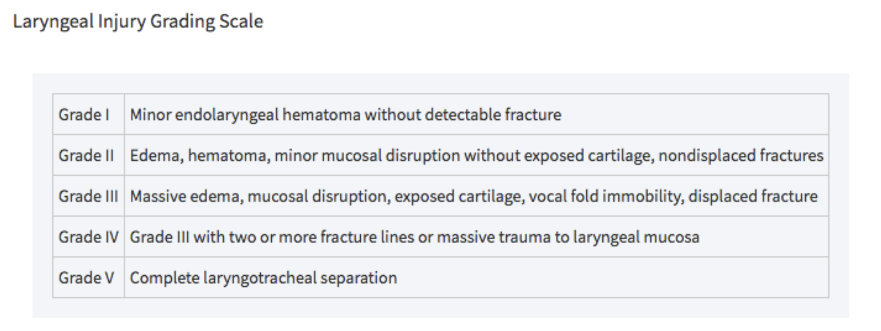
          • Grades III, IV, and V laryngotracheal injuries as defined by Schaefer and Brown’s classification system require OR

    Tintinalli 2018

          • Involve consultants early: trauma surgery, neurosurgery, vascular surgery, neurology, otolaryngology
    • Cervical spine/ spinal cord injury
      • See chapter for spinal trauma

    Disposition

    • Admit symptomatic patients to monitored setting
    • Given delayed symptoms, consider monitoring patients who are asymptomatic on arrival
      • Serial exams for worsening dyspnea, dysphonia, stridor, drooling, bruits, focal neuro deficits
      • Only discharge after ruling out airway threat, neurological deficit, vascular injury, or suicidal/ homicidal ideation
      • Monitor asymptomatic patients on home anticoagulation in ED for at least 6 hours from trauma to rule out delayed neck hematoma
    • Social work and/or psychiatry for patients in whom you suspect suicide risk or domestric violence, look for other signs of self harm

    Take Home Points

    • Aggressive early airway management for unconscious patient, evidence of airway compromise including voice change, dyspnea, neurological changes, or pulmonary edema
    • Involve consultants early: trauma surgery, neurosurgery, vascular surgery, neurology, otolaryngology
    • Victims of blunt cerebral vascular injury may present completely asymptomatic but develop delayed neurological symptoms; close observation and monitoring is recommended especially for patients on home anticoagulation
    • Remember to evaluate for concomitant injuries
    • Psychiatric evaluation for all attempted suicides

    References

    • Bromberg, William. et al. Blunt Cerebrovascular Injury Practice Management Guidelines: The Eastern Association for the Surgery of Trauma. J Trauma. 68 (2): 471-7, Feb 2010.
    • Cothren CC, Moore EE, Biffl WL, et al. Anticoagulation is the gold standard therapy for blunt carotid injuries to reduce stroke rate. Arch Surg. 2004;139:540–545; discussion 545–546.
    • Joshua AA. Neck Trauma, Blunt, Anterior. In: Schaider J, Barkin R, Hayden S, Wolfe R, Barkin A, Shayne P, Rosen P. Rosen and Barkin’s 5-Minute Emergency Medicine Consult. 5th Edition. Philadelphia, PA: Wolters Kluwer; 2015; 738-739.
    • Tintinalli, J., Stapczynski, J. Stephan, editor, Ma, O. John, editor, Yealy, Donald M., editor, Meckler, Garth D., editor, & Cline, David, editor. (2018). Tintinalli’s emergency medicine : A comprehensive study guide (9th ed.).
    • Walls, R., Hockberger, Robert S., editor, & Gausche-Hill, Marianne, editor. (2018). Rosen’s emergency medicine : Concepts and clinical practice (Ninth ed.).
    • Advanced trauma life support. (2018). 10th ed. Chicago, IL: American College of Surgeons.

    Special thanks to Sana Maheshwari, MD

    NYU Bellevue Emergency Medicine Residency PGY3


    Read More

    Episode 172.0 – Ankle Sprains Nov 04, 2019
    Show notes

    We dissect one of the most common injuries we see in the ER -- ankle sprains

    Hosts:
    Brian Gilberti, MD
    Audrey Bree Tse, MD

    https://media.blubrry.com/coreem/content.blubrry.com/coreem/Ankle_Sprains.mp3 Download 3 Comments Tags: Orthopedics

    Show Notes

    Background

    • Among most common injuries evaluated in ED
    • A sprain is an injury to 1 or more ligaments about the ankle joint
    • Highest rate among teenagers and young adults
      • Higher incidence among women than men
    • Almost a half are sustained during sports
    • Greatest risk factor is a history of prior ankle sprain

    Anatomy

    • Bone: Distal tibia and fibula over the talus → constitutes the ankle mortise
    • Aside from malleoli, ligament complexes hold joint together
      • Medial deltoid ligament
      • Lateral ligament complex
        • Anterior talofibular ligament
          • Most commonly injured
          • Weakest
          • 85% of all ankle sprains
        • Posterior talofibular ligament
        • Calcaneofibular ligament
      • Syndesmosis

    Mechanism of Injury

    • Lateral ankle sprains
      • Most common among athletes
      • ATFL most commonly injured
        • Combined with CFL in 20% of injuries
      • 2/2 inversion injuries
    • Medial ankle sprains
      • Less common than lateral because ligaments stronger and mechanism less frequent
        • More likely to suffer avulsion fracture of medial malleolus than injure medial ligament
      • 2/2 eversion +/- forced external rotation
      • Typically landing on pronated foot -> external rotation
    • High Ankle sprains
      • Syndesmotic injury
      • More common in collision sports (football, soccer, etc)
    • Grade I
      • Mild
      • Stretch without “macroscopic” tearing
      • Minimal swelling / tenderness
      • No instability
      • No disability associated with injury
    • Grade II
      • Moderate
      • Partial tear of ligament
      • Moderate swelling / tenderness
      • Some instability and loss of ROM
      • Difficulty ambulating / bearing weight
    • Grade III
      • Severe
      • Complete rupture of ligaments
      • Extensive swelling / ecchymosis / tenderness
      • Mechanical instability on exam
      • Inability to bear weight

    Examination

    • Beyond visual inspection for swelling, ecchymoses, abrasions, or lacerations
      • Palpation
      • Pain when palpating ligament is poorly specific but may indicate injury to structure
      • Check sites for Ottawa ankle rules to evaluate if there may be an associated fracture with injury
        • Posterior edge or tip of lateral malleolus (6 cm)
        • Posterior edger or tip of medial malleolus (6 cm)
        • Base of fifth metatarsal
        • Navicular bone
      • Acute ATFL rupture / Grade III Sprain
        • 90% chance of this injury if hematoma and localized tenderness with palpation present on exam over this ligament
        • Anterior drawer test
          • Assess for anterior subluxation of talus from the tibia
            • Ankle in relaxed position, distal extremity is stabilized with one hand while the other cups the heel to apply anterior force
          • Compare to contralateral side
          • Difficult to determine if there is an acute rupture at this point and may be more easily diagnosed in subacute phase (4-5 days after injury)
          • Ability to perform exam adequately limited by pain, swelling and potential muscle spasm
        • Talar tilt test
          • If applying inversion force to ankle and there is excessive mobility → calcaneofibular ligament
        • Thompson test
          • Can be performed if there is concern for concomitant Achilles tendon injury
      • Do not miss a Maisonneuve fracture by palpating proximally about the fibular ahead as forces may be transmitted through the syndesmosis
        • Squeeze test – pressure just proximal to ankle
          • If elicits pain → concern for syndesmotic injury

    Diagnostics

    • X-rays indicated if unable to rule out using Ottawa Ankle Rules
      • Sn (Up to 99.6) (one of the best validated tools we use in the ER)
      • May have trouble applying rule if there is question of patients ability to sense pain (diabetic neuropathy), in which case obtain radiographs

    Treatments

    • RICE
      • Crutch train so they can be weight bearing a tolerated
      • Ideally initiate within first 24 hours of injury
      • Ice 15-20 minutes q2-3h over the first 48 hours or until swelling improves
    • NSAIDs
      • Topical and PO are better than placebo
      • We do not know if PO is superior to topical NSAIDs
    • Early mobilization / Functional Rehab (sample patient instructions here)
      • Work to restore range of motion, strength, proprioception
      • For Grade I and II, can begin as soon as the patient can tolerate and ideally within 1 week of the injury
      • Patients return to work sooner, decreased chronic instability, less recurrent injuries
      • Dorsiflexion, plantarflexion, and perform foot circles as well as toe curls, inversion and eversion as tolerated
      • Proprioception
        • Balancing on wobble board
      • Continue exercises until patient is able to return to activities at full capacity, without pain
    • Immobilization
      • High re-injury rates and important to protect against this
      • Grade I
        • No immobilization required
        • +/- Ace wrap
      • Grade II
        • Aircast brace
        • Ensure patient understands that they should still partake in rehabilitation exercises
      • Grade III
        • Data conflicts
        • RCT, multicenter study comparing aircast brace, compression bandage, Bledsoe immobilization boot and below-knee cast for 10 days
          • Ankle function at 3 months
            • Cast group had most improvement
            • No difference at 9 months in function or complications
        • May be institution-dependent and a cast can be offered initially

    Prognosis

    • Acute inflammation → reduction in swelling → development of new tissue → strengthening of tissue
    • Return of basic function, though limited, occurs over 4-6 weeks depending on severity of sprain
    • Try to limit strain put on joint (no heavy lifting, walking on uneven surfaces, try to limit standing while at work)
    • Follow up:
      • If pain or instability does not improve over 4-6 weeks
      • Grade III sprains
      • Medial ankle sprains (may have underlying fracture that was undetected in ED on XR)
      • Syndesmosis injuries (protracted recovery course)
      • Injuries associated with fractures or dislocation / subluxation

    Read More

    Episode 171.0 – Vaping Associated Lung Injury Oct 21, 2019
    Show notes

    An overview of Vaping Associated Lung Injury (VALI)

    Hosts:
    Audrey Bree Tse, MD
    Larissa Laskowski, DO
    Brian Gilberti, MD

    https://media.blubrry.com/coreem/content.blubrry.com/coreem/Vaping_Associated_Lung_Injury.mp3 Download 2 Comments Tags: Pulmonary, Toxicology

    Show Notes

    Why this matters

    • As of Oct 15, vaping has been associated with acute lung injury in over 1400 people
    • 33 deaths have been confirmed in 24 states
    • 70+% of those with VALI are young men
    • A large number of patients are requiring ICU/ intubation/ ECMO

    4 main ingredients in solvent

    • +/- Flavor additives
    • +/- Nicotine or THC (Tetrahydrocannabinol)
    • Propylene Glycol (PG)
    • Vegetable Glycerin (VG)

    CDC definition of VALI (Vaping Associated Lung Injury)

    1. Using an e-cigarette (“vaping”) or dabbing* in 90 days prior to symptom onset AND
    2. Pulmonary infiltrate, such as opacities, on plain film chest radiograph or ground-glass opacities on chest CT AND
    3. Absence of pulmonary infection on initial work-up.
    4. No evidence in the medical record of alternative plausible diagnoses (e.g., cardiac, rheumatologic, or neoplastic process).

    *Dabbing allows the user to ingest a high concentration of THC. Butane Hash Oil (BHO), an oil or wax-like substance extracted from the marijuana plant, is placed on a “nail” attached to a specialized glass bong called a “rig.” A blow torch is used to heat the wax, which produces a vapor that can then be inhaled to supposedly produce an instantaneous effect.

    Pathophysiology

    • At present, no single compound or ingredient has emerged as the cause, and there may be more than one cause
    • The only common thread among the cases is that ALL patients reported using e-cig or vaping products
    • Leading potential toxins:
      • Vaping products containing THC concentrates: most cases are linked to THC concentrates that were either purchased on the street or from other informal sources (meaning not from a dispensary)
      • Vitamin E acetate: nutritional supplement safe when ingested or applied to the skin (but likely not when inhaled) has been found in nearly all product samples of NY state cases of suspected VALI
        • vitamin E acetate is NOT an approved additive at least by NYS Medical Marijuana program
    • Other potential toxins:
      • IT CANNOT BE UNDERSTATED that a small percentage of persons w/ VALI have reported exclusive use of nicotine-containing vape products, such as JUUL; as such, we must consider the potential toxicity of standard e-liquid or vape juice
      • Flavor additives, that exists as chemical aldehydes: irritating and potentially damaging to lung tissue
      • PG/VG: shown not only to break down to formaldehyde which is a known carcinogen, but also to produce lipoid pneumonia in rat lungs
      • Some devices are easily manipulated to increase the capacity to produce vapor; increasing these settings may impact heating temperature, metabolic breakdown, and release of microscopic metal particles
      • Lungs are multifunctional, including serving as an immune organ: lungs cleave proteins of all of the bacteria, viruses and other pathogens we are exposed to and inhale daily
        • human studies on those that are chronic e-cig users or vapers have revealed that these products are shifting the balance of proteases and antiproteases in our lungs such that the proteases are destroying native lung tissue similar to how traditional cigarettes cause COPD
    • Many potential reactions:
      • NEJM article in references: details four radiographic phenotypes essentially reflecting different pathologic changes

    Long-term Effects

    • Long term effects are unknown (some pts have required home oxygen on discharge)
    • Risk for recurrence or relapse, especially if repeat exposure

    Presentation

    • 95% of pts have had pulmonary sxs (cough, cp, dyspnea)
    • 77% of pts have had GI sxs (abd pain, n/v/d)
    • 85% of pts w/ constitutional sxs (f/c, weight loss)
    • 57% w/ hypoxia (O2 < 95%)
    • Unfortunately auscultation has been unreliable and poorly sensitive

    Workup

    • There is no specific test or marker for dx, so VALI is still considered a dx of exclusion
      • Labs:
        • CBC
        • ESR/CRP (93% w/ elevated ESR)
        • LFTs (50% w/ transaminitis)
        • ABG: hypoxia
      • Imaging:
        • CXR: typically shows bilateral infiltrates, although not always and there have even been some cases w/ unremarkable chest XR (so high degree of clinical suspicion in any person p/w hypoxia)
        • CT: ground glass opacities, typically bilaterally

    Management

    • Dispo:
      • 96% of cases required hospitalization
        • Any pt w/ hypoxia, respiratory distress, or comorbidities
      • Outpatient only if: no hypoxia or respiratory distress, reliable followup within 48h and good social support (keep in mind that some patients w/ mild symptoms of first presentation deteriorated rapidly within 48h)
    • Empiric treatments for pneumonia inc abx, antivirals
    • Steroids (methylpred 60mg q6h, based on how index cases in Illinois were managed)
      • Case reports have documented improvement
      • Mechanism: blunting of inflammatory response
    • Aggressive supportive care

    Special Thanks To:

    Dr. Larissa Laskowski, DO

    Ronald O. Perelman Department of Emergency Medicine at NYU Langone Health, NYC Health + Hospitals/ Bellevue

    New York City Poison Control Center

    References:

    Outbreak of Lung Injury Associated with E-Cigarette Use, or Vaping. https://www.cdc.gov/tobacco/basic_information/e-cigarettes/severe-lung-disease.html

    Carlos WG, Crotty Alexander LE, Gross JE, Dela Cruz CS, Keller JM, Pasnick SP, Jamil S. Vaping-associated Pulmonary Illness (VAPI). Public Health Information Series. Am J Respir Crit Care Med Vol. 200, 13-15, 2019. www.atsjournals.org/doi/pdf/10.1164/rccm.2007P13

    Henry TS, Kanne JP, Kilgerman SJ. Images of Vaping-Associated Lung Disease — Correspondence. N Engl J Med. 2019 Oct 10; 381;15.

    Layden JE, Ghana I, Pray I, Kimball A, Layer M, Tenforde M, Navon L, Hoots B, Salvatore PP, Elderbrook M, Haupt T, Kanne J, Patel MT, Saathaff-Huber L, King BA, Schier JG, Mikosz CA, Meiman J. Pulmonary Illness Related to E-Cigarette Use in Illinois and Wisconsin – Preliminary Report. N Engl J Med. 2019 Sep 6. doi: 10.1056/NEJMoa1911614. [Epub ahead of print]. https://www.ncbi.nlm.nih.gov/pubmed/31491072?dopt=AbstractPlus

    Siegel DA, Jatlaoui TC, Koumans EH, et al. Update: Interim Guidance for Health Care Providers Evaluating and Caring for Patients with Suspected E-cigarette, or Vaping, Product Use Associated Lung Injury — United States, October 2019. MMWR Morb Mortal Wkly Rep 2019;68:919–927. DOI: http://dx.doi.org/10.15585/mmwr.mm6841e3external icon.

    https://www.health.ny.gov/press/releases/2019/2019-09-05_vaping.htm


    Read More

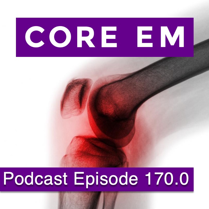
    Episode 170.0 – Septic Arthritis Sep 23, 2019
    Show notes

    An overview of septic arthritis.

    Hosts:
    Audrey Bree Tse, MD
    Brian Gilberti, MD

    https://media.blubrry.com/coreem/content.blubrry.com/coreem/Septic_Arthritis.mp3 Download One Comment Tags: Infectious Diseases, Orthopedics

    Show Notes

    Episode Produced by Audrey Bree Tse, MD

    Background

    • Bacteria enters the joint by hematogenous spread due to absence of basement membrane in synovial space from invasive procedures, contiguous infection (e.g. osteomyelitis, cellulitis), or direct inoculation (e.g. plant thorns, nails)
      • WBCs migrate into joint → acute inflammatory process → synovial hyperplasia, prevents new cartilage from forming, pressure necrosis on surrounding joint, purulent effusion
    • Why do we care?
      • irreversible loss of function in up to 10% & mortality rate as high as 11%
      • Cartilage destruction can occur in a matter of hours
      • Complications include bacteremia, sepsis, and endocarditis

    Etiology

    • Risk factors: extremes of age, RA, DJD, IVDA, endocarditis, GC, immunosuppression, trauma, or prosthesis
    • Organisms:
      • Staph: staph aureus (most common), MRSA, Staph epidermis
      • N gonorrhea: young healthy sexually active adults
      • Strep: group A & B
      • GNRs: IVDA, diabetics, elderly
      • Salmonella: sickle cell disease
      • Cutibacterium acnes: prosthetic shoulder infection
      • Consider mycobacterial & fungal in more indolent courses

    Presentation

    • Typically a single, warm, erythematous, tender joint (#1: knee (50% of cases) → hip, shoulder, ankle)
      • *Any joint can be involved!
      • IVDA can involve sacroiliac, costochondral, & sternoclavicular joints
      • Classic teaching: very painful with ROM, but this is not always present!
      • Joint usually held in position of maximum joint volume
      • Prosthetic joints may have less pain than expected for a septic joint given changed anatomy and disrupted nerve endings
    • In 10-20% of cases, can see polyarticular involvement
      • GC typically monoarticular but commonly polyarticular
    • Often have fever & separate infection as well (only see fever in ~60% of cases)

    Diagnostics

    Arthrocentesis:

    • Gold standard
    • Tap joint even if acceptable ROM: septic joints can have normal motion so it does not exclude the diagnosis!
    • Use ultrasound if possible
    • Relative contraindications: overlying cellulitis (risk of seeding joint) or severe coagulopathies (weigh risk of creation or worsening of iatrogenic hemarthrosis)
    • Keep in mind that a “dry tap” may occur due to incorrect needle placement, absent/ minimal joint effusion, ort mechanical obstruction
    • Note: talk to ortho colleagues if prosthesis present prior to performing arthrocentesis
      • Ortho team may want to perform the arthrocentesis themselves because scar tissue formation and altered anatomic relationships make the procedure more challenging
      • Usually want to perform washout in OR plus/ minus antibiotic spacer
    • Send fluid for protein, glucose, cell count with differential, gram stain, culture, and crystals
      • Often see decreased glucose and elevated protein
      • The presence of crystals does not rule out septic arthritis
      • No clear number of synovial WBCs to define septic arthritis, but in general: >30 to 50K/ mm3 synovial WBCs with PMN predominance (>75%) seen in septic arthritis
      • A 2011 meta-analysis suggests +LRs of 4.7 (95% CI = 2.5 to 8.5) and +LR of 13.2 (95% CI = 3.6 to 51.1) for a sWBC count of >50L × 109 or >100K, respectively
      • Use the synovial WBC count plus the whole clinical picture to rule in or out the diagnosis of septic arthritis (do not use the synovial WBC in isolation)
      • Different threshold for prosthetic joints: WBC > 1100 or >64% PMNs = septic arthritis
    • Gram stains only identify causative organisms 1/3 of the time
    • Culture negative arthrocentesis can be seen in cases where abx have been given prior to arthrocentesis, or in TB/ brucella/ nocardia/ other indolent organisms like fungi

    Labs:

    • No studies have demonstrated an acceptable sensitivity or overall diagnostic accuracy of peripheral WBC count for SA, but usually see leukocytosis with left shift
    • ESR and CRP are reasonably sensitive but there is no cutoff that significantly increases or decreases the pretest probability
    • UA, urine cultures, blood cultures: send even if no fever
      • Blood cultures are positive in 50-70% of nonGC SA
    • If GC suspected, do GC NAAT from throat/ rectal/ urethral/ cervical discharge

    Imaging:

    • XRs: effusion, baseline status of joint, contiguous osteomyelitis, fractures, foreign body
    • US: effusion
    • CT, MRI: not really used in ED

    Differential

    • Viral arthritis
    • RA
    • gout/ pseudogout
    • HIV associated arthritis
    • Reactive arthritis
    • Lyme
    • Osteo
    • Septic bursitis
    • Trauma

    Treatment

    • Septic arthritis is an orthopedic emergency!
      • Needs IV abx + often washout of the joint
    • Hold abx as much as possible prior to tap unless pt is unstable or tap cannot be performed easily
    • Initiate empiric IV antibiotic therapy prior to definitive cultures based
    • Transition to organism-specific antibiotic therapy once culture sensitivities result
    • Start empiric abx based on gram stain if available (in non-=GC SA, grain stain is positive in 50% of cases), age group, & risk factors
      • Empiric abx: Vancomycin 15mg/kg q12h (to cover MRSA) + cefepime 2gr IV q8h (to cover gram-negatives)
      • If gram stain with GPC = Vancomycin 15mg/kg q12h
      • If gram stain with GN diplococci = ceftriaxone 1gr IV q24h + Azithromycin 1gr q24h
      • If gram stain with GN rods = cefepime 2gr IV q8h
      • If penicillin allergy: ciprofloxacin 500mg q12h or aztreonam 2gr q8h
      • No need to cover anaerobes unless human/dog/cat bite (then use Unasyn to cover eikenella, pastereulla, capnocytophaga, anaerobes, etc.)
    • They usually need antibiotics for 2-6 weeks: 2 weeks for strep, up to 6 weeks if S aureus
    • Pain control: consider moderately flexed splinting
    • Admit all patients with suspected septic arthritis until SA is ruled out, abx, monitoring, likely operative intervention

    Take-Home Points

    • Patients may present with either a single affected joint or polyarticular; they may or may not have a fever
    • Have a high index of suspicion for SA, and a low threshold to tap: pts do not necessarily present w/ “classic” findings and it is difficult to distinguish SA from crystal arthropathy
    • ESR, CRP, serum WBC are not definitive diagnostic tools for septic arthritis
    • There is no exact cutoff for synovial WBCs for diagnosis: use whole clinical picture & keep 50K in mind for native joints, and >1100 for prostheses
    • Treat with empiric abx after tap then narrow accordingly, & admit all patients with septic arthritis
    • Involve your ortho colleagues early especially for prosthesis

    References

    Carpenter CR, Schuur JD, Everett WW, et al. Evidence-based diagnostics: Adult septic arthritis. Acad Emerg Med. 2011;18:781-796.

    Jones D, Clements C. Physical exam and bloodwork do not adequately differentiate infectious from inflammatory arthritis. In: Mattu A, Chanmugam A, Swadron S, Woolridge D, Winters M. Avoiding Common Errors in the Emergency Department. 2nd Edition. Philadelphia, PA: Wolters Kluwer; 2017; 412-414.

    Kazzi A, Zaghrini E. Septic Arthritis. In: Schaider J, Barkin R, Hayden S, Wolfe R, Barkin A, Shayne P, Rosen P. Rosen and Barkin’s 5-Minute Emergency Medicine Consult. 5th Edition. Philadelphia, PA: Wolters Kluwer; 2015; 102-103.

    Osmon D, Berbari E, Berendt A, Lew D, Zimmerli W, Steckelberg J, Rao N, Hanssen A, Wilson W.

    Diagnosis and Management of Prosthetic Joint Infection: Clinical Practice Guidelines by the Infectious Diseases Society of America, Clinical Infectious Diseases, Volume 56, Issue 1, 1 January 2013, Pages e1–e25, https://doi.org/10.1093/cid/cis803

    Mlynarek C, Sullivan A. Arthrocentesis Tips. In: Mattu A, Chanmugam A, Swadron S, Woolridge D, Winters M. Avoiding Common Errors in the Emergency Department. 2nd Edition. Philadelphia, PA: Wolters Kluwer; 2017; 684-686.

    Purcell D, Terry B, Sharp B. Joint Arthrocentesis. In: Purcell D, Chinai S, Allen B, Davenport M. Emergency Orthopedics Handbook. 1st Edition. Cham, Switzerland: Springer; 2019; 87-104.

    Sheth U, Moore D. Septic Arthritis — Adult. OrthoBullets. [https://www.orthobullets.com/trauma/1058/septic-arthritis–adult]. Updated 1/9/19. Accessed 8/2/19.

    A special thanks to our Infectious Diseases Editor:

    Angelica Cifuentes Kottkamp, MD

    Infectious Diseases & Immunology

    NYU School of Medicine

    A special thanks to our Orthopedics Editor:

    Daniel Purcell, MD

    Emergency Medicine

    NYU Langone Brooklyn


    Read More

    Episode 169.0 – Febrile Seizures Aug 26, 2019
    Show notes

    A look at the most common type of seizures in the young pediatric population.

    Hosts:
    Brian Gilberti, MD
    Audrey Bree Tse, MD

    https://media.blubrry.com/coreem/content.blubrry.com/coreem/Febrile_Seizures.mp3 Download Leave a Comment Tags: Pediatrics

    Show Notes

    Background

    • The most common type of seizure in children under 5 years of age
    • Occur in 2-5% of children
    • In children with a fever, aged 6 months to 5 years of age, and without a CNS infection
    • Risk Factors
      • 4 times more likely to have a febrile seizure if parent had one
      • Also increase in risk if siblings or nieces / nephews had one
    • Common associated infections
      • Human Herpesvirus 6
      • Human Herpesvirus 7
      • Influenza A & B
    • Simple Febrile Seizure
      • Generalized tonic-clonic activity lasting less than 15 minutes in a child 6 months to 5 years of age
    • Complex Febrile Seizure
      • Lasts longer than 15 minutes, occurs in a child outside of this age range, are focal, or that recur within a 24-hour period.

    Diagnostics / Workup

    • Gather thorough history and perform thorough physical exam
    • Most cases will not require labs, imaging or EEG
    • If e/o meningitis, perform LP
    • AAP suggests considering LP in:
      • Children 6-12 months who are not immunized for H flu type B or strep pneumo
      • Children who had been on antibiotics
    • For complex seizures, clinician may have a lower threshold for obtaining labs
      • Hyponatremia is more common in this group than in the general population.
      • LPs are more commonly done by providers, but these are low yield with one study showing bacterial meningitis being diagnosed in just 0.9% (Kimia 2010), all of whom did not have a normal exam or negative cultures.
      • Neuroimaging is also exceedingly low yield if the patient returns to baseline (Teng 2006)
      • One study that showed that the duration of complex febrile seizure, being greater than 30 minutes, was associated with a higher incidence of bacterial meningitis. (Chin 2005)
      • Of they have history and exam concerning for meningitis, they should get an LP
      • If they look dehydrated or edematous, you would have more of a reason to get a chemistry

    Treatment

    • Benzodiazepine if seizure lasted for >5 minutes, either IV or IN
    • Supportive care
      • Tylenol or motrin if febrile
      • Fluids if signs of dehydration
    • Antipyretics “around the clock”
      • A majority of data show no benefit in preventing recurrence of seizure
      • One study (Murata 2018) found that giving tylenol q6h at 10 mg/kg for the first 24 hours following the initial seizure decreased the rate of recurrence when compared to children who did not receive antipyretics.
        • NNT here was 7
        • Questionable whether we can generalize these findings from a single ED in Japan.
      • No role for antiepileptics

    Prognosis

    • High rate of recurrence (~1/3) within 1 year of initial seizure
    • Risk increases for
      • Younger age at which they had initial seizure
      • Lower temperature at which they had seizure
    • If initial febrile seizure was prolonged, more likely that the next will be prolonged
    • 1-2% develop epilepsy for simple febrile seizure, slightly above risk of general population
    • 5-10% develop epilepsy for complex febrile seizure
    • Follow up with PMD
    • Generally, peds neuro follow up is not necessary

    References

    Chin RF, Neville BG, Scott RC. Meningitis is a common cause of convulsive status epilepticus with fever. Arch Dis Child. 2005;90(1):66-9.

    Kimia A, Ben-Joseph EP, Rudloe T, Capraro A, Sarco D, Hummel D, et al. Yield of lumbar puncture among children who present with their first complex febrile seizure. Pediatrics. 2010;126(1):62-9.

    Murata S, Okasora K, Tanabe T, Ogino M, Yamazaki S, Oba C, et al. Acetaminophen and Febrile Seizure Recurrences During the Same Fever Episode. Pediatrics. 2018;142(5).

    Patel N, Ram D, Swiderska N, Mewasingh LD, Newton RW, Offringa M. Febrile seizures. BMJ. 2015;351:h4240.

    Pavlidou E, Panteliadis C. Prognostic factors for subsequent epilepsy in children with febrile seizures. Epilepsia. 2013;54(12):2101-7.

    Stapczynski, J. S., & Tintinalli, J. E. (2016). Tintinalli’s emergency medicine: A comprehensive study guide, 8th Edition. New York: McGraw-Hill Education.

    Subcommittee on Febrile S, American Academy of P. Neurodiagnostic evaluation of the child with a simple febrile seizure. Pediatrics. 2011;127(2):389-94.

    Teng D, Dayan P, Tyler S, Hauser WA, Chan S, Leary L, et al. Risk of intracranial pathologic conditions requiring emergency intervention after a first complex febrile seizure episode among children. Pediatrics. 2006;117(2):304-8.

    Warden CR, Zibulewsky J, Mace S, Gold C, Gausche-Hill M. Evaluation and management of febrile seizures in the out-of-hospital and emergency department settings. Ann Emerg Med. 2003;41(2):215-22.

    A special thanks to our editors:

    Michael A. Mojica, MD

    Director, Pediatric Emergency Medicine Fellowship
    Bellevue Hospital Center

    Christie M. Gutierrez, MD

    Pediatric Emergency Medicine Fellow
    Columbia University Medical Center
    Morgan Stanley Children’s Hospital
    New York Presbyterian


    Read More

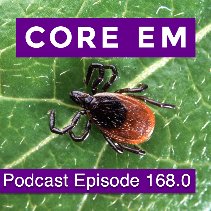
    Episode 168.0 – Lyme Disease Jul 30, 2019
    Show notes

    A review for the emergency physician of this common tick-borne illness.

    Hosts:
    Audrey Bree Tse, MD
    Brian Gilberti, MD

    https://media.blubrry.com/coreem/content.blubrry.com/coreem/Lyme_Disease.mp3 Download Leave a Comment Tags: Infectious Diseases

    Show Notes

    Episode Produced by Audrey Bree Tse, MD

    Background

    • Most common tick-born illness in North America
    • Endemic in Northeast, Upper Midwest, northwest California
    • 80% to 90% in summer months

    Pathophysiology

    • Ixodes tick (deer tick) has a 3-stage life cycle (larvae, nymph, adult) & takes 1 blood meal per stage
    • Deer tick feeds on an infected wild animal (infected with spirochete Borrelia burgodrferi) then bites humans
    • On humans, they typically move until they encounter resistance (e.g. hairline, waistband, elastic, skin fold). It takes 24-48 hrs for B. Burgdorferi to move from the tick to the host
    • Pathogenesis: organism induced local inflammation, cytokine release, autoimmunity
    • No person to person transmission

    Clinical Presentation

    Stage 1: Early

    • Symptom onset few days to a month after tick bite
    • Erythema migrans rash: bulls eye rash seen in more than 90% of patients with Lyme disease (Irregular expanding annular lesion(s))
    • Regional adenopathy, intermittent fevers, headache, myalgias, arthralgia, fatigue, malaise

    Stage 2: disseminated/ secondary

    • Days to weeks after tick bite
    • Intermittent fluctuating sx that eventually resolve
    • Triad of aseptic meningitis, cranial neuritis, and radiculoneuritis: bell palsy most common
    • Cardiac symptoms: tachycardia, bradycardia, AV block, myopericarditis

    Stage 3: tertiary/ late

    • Symptoms occur >1 year after tick bite
    • Acrodermatitis chronic atrophicans: Atrophic lesions on extensor surfaces of extremities (resembles scleroderma)
    • Monoarthritis, oligoarthritis (knee > shoulder > elbow)
    • GI: Hepatitis, RUQ pain
    • Ocular: keratitis, uveitis, iritis, optic neuritis
    • Neurological: Chronic axonal polyneuropathy or encephalopathy

    Chronic Lyme disease (versus well-accepted Lyme disease sequelae):

    • Continuation of symptoms after antibiotics
    • Current recommendation for management is supportive care only

    Pediatric considerations:

    • More likely to be febrile than adults
    • Facial palsy accompanied by aseptic meningitis in 1/3
    • Untreated kids can develop keratitis
    • Excellent prognosis if appropriately treated

    History

    • Travel, camping, woods, playing under leaves or in wood piles
    • Living in endemic area (Northeastern area: Maine to Virginia; upper Midwestern: Wisconsin, Minnesota; Northwest California)
    • Endemic in Northern Europe and Eastern Asia as well
    • History of tick bite (- 30-50% of patients recall tick bite)
    • Flu like illness in summer
    • Rash: https://www.cdc.gov/lyme/signs_symptoms/rashes.html
    • Joint complaints
    • Cardiac complaints
    • Neurologic complaints
    • Careful search for tick

    Diagnosis

    Labs

    • CBC (leukocytosis, anemia, thrombocytopenia)
    • ESR: most common lab abnormality (>30 mm/hr)
    • Chem 7
    • LFTs: commonly elevated especially GGT
    • Cultures not typically indicated
    • LP when meningeal signs (CSF: pleocytosis, elevated protein, CSF spirochete ABs). LP function is more to rule out other etiologies of meningitis rather than diagnose Lyme meningitis given that lyme PCR and lyme Ab index are not very accurate.

    Serological Testing

    • Serological testing is not always warranted because of the very high incidence of false positive results
    • Serologies are not useful in acute phase (<30 days of infection) because they are negative; it takes several weeks to develop enough antibodies for either test below (ELISA or Western Blot)
    • Acute Lyme is a clinical diagnosis and does not need laboratory testing, especially in endemic areas such as NY
    • If pretest probability is high (symptoms consistent with Lyme + epidemiological background), say patients with CN palsy, meningitis, carditis, or migratory large joint arthritis, then serologies can be very helpful
    • Do not test if patients in endemic areas with potential tick exposure present with EM — just treat with antibiotics
    • Do not test if patients in endemic areas present with no history of tick exposure or only nonspecific symptoms
    • Test if you have high suspicion of lyme without EM
    • PCR is highly specific and sensitive but not available for routine use. There are two tests you need to use together:

    1) ELISA: this detects antibodies to lyme bacteria (borrelia burgdorferi) in your blood, BUT it can’t distinguish between borrelia and similar bacteria (even sometimes normal flora that lives in you). In addition, IgM response takes 1-2 weeks while IgG response takes 2-4 weeks.

    If ELISA is positive or equivocal, then you move onto the:

    2) Western blot test: this looks for antibodies not to the whole organism, but to the basic building blocks of the lyme bacteria — the individual proteins, BUT many types of bacteria use the same building blocks. So the CDC says that the Western Blot test must detect IgG antibodies to 5 out of the 10 proteins.

    See figure 2: https://www.ncbi.nlm.nih.gov/pmc/articles/PMC4918152/

    • Two-tiered testing has sensitivity between 70-100% and specificity ~95% in late stages
    • Interpretation of Lyme serologies should be done by an ID specialist because they can be confusing and can lead to wrong conclusions if unfamiliar with them
    • NYC is an endemic region where 5% of the population can have a positive without symptoms!
    • If somebody who HAD Lyme disease but successfully treated it with doxycycline tested themselves years later, they could still have the antibodies and therefore it would look like they still had Lyme disease (despite being cured)
    • Positive serology or previous Lyme disease not ensure protective immunity

    Other tests:

    • Arthrocentesis for acute arthritis: elevated cryoglobulin
    • XRs: may show soft tissue, cartilaginous, osseous changes
    • ECG

    Differential Diagnosis

    • Tick-borne diseases: Rocky Mountain Spotted fever, tularemia, relapsing fever, Colorado tick fever, tick-bite paralysis, babesiosis, anaplasmosis, powassan virus
      • Remember that doxycycline covers anaplasmosis and lyme but not babesiosis, which requires Atovaquone
    • Rheumatic fever (usually presents with erythema marginatum rash, valvular involvement rather than heart block, TM joint arthritis)
    • Viral meningitis
    • Septic arthritis
    • Syphilis
    • Parvovirus B19
    • Infectious endocarditis
    • Juvenile rheumatoid arthritis
    • Reiter syndrome
    • Brown recluse spider bite
    • Fibromyalgia
    • Chronic fatigue syndrome

    Treatment

    • Remove tick: disinfect site then with blunt instrument, grasp tick proximal to skin and pull upward with gentle constant traction. Mouthparts will release after about a minute. If residual mouthparts are left in skin, leave them alone to avoid infection (they will extrude from skin naturally over time). Since ticks that have not attached or are moving on the skin cannot transmit Lyme, they can just be brushed off.
    • NS IVF bolus, supportive care
    • Cardiac monitoring, temporary pacemaker for heart block
    • Beware Jarisch Herxheimer reaction: worsening of sx a few hours after treatment initiated
    • Aspirin for cardiac involvement, NSAIDs for arthralgias/ arthritis

    Prophylaxis:

    • Per the IDSA, give a single dose of 200 mg PO doxycycline to patients who meet all of the following criteria:
      • Deer tick has been attached for 36 hours or more (the rationale for time of attachment relates to the fact that the spirochetes live in the tick’s gut so they need a long time to multiply and travel to the salivary glands (event that’s triggered by a blood meal) and later overcome the salivary gland (which only a few do) and finally reach the patient’ skin
      • Prophylaxis can be provided within 72 hours of tick removal
      • Local rate of B. Burgdorferi infection in ticks exceeds 20% (in the northeast USA, the prevalence of infected ticks is between 15-20%)
      • Doxycycline can be used (children >8 years old, non-pregnant females)
    • A 2001 study examined doxycycline vs placebo prophylaxis. A single dose of 200 mg of oral doxycycline or placebo was given to persons presenting within 72 hours of removal of an I scapularis tick. One of 235 persons in the doxycycline group developed erythema migrans (EM) versus 8 of 247 in the placebo group, for treatment efficacy of 87% (95% CI, 25%–98%; P<0.04) (9).
    • Reasonable alternative strategy: monitor for EM or other signs of infection then initiate treatment if they develop Lyme disease (excellent outcomes in patients treated during early EM stage of disease)

    Antibiotics:

    • Antibiotics can speed resolution of arthritis and cardiac conduction delays, but not necessarily facial palsy
    • Doxycycline has the best bioavailability and CNS penetration
    • Always check with your ID colleagues to determine appropriate duration of treatment in more serious cases of Lyme disease
    • Stage 1:
      • Amoxicillin (500 mg PO TID) or cefuroxime (500 mg PO BID) or doxycycline (100 mg PO BID; > 8 years old & not pregnant) x 21 days; azithromycin (500 mg PO qday x 14-21 days)
      • IV therapy in pregnant patients
    • Stage 2:
      • PO antibiotics for isolated Bell palsy and mild involvement
      • Amoxicillin with probenecid (500 mg PO TID) x 30 days or doxycycline (100 mg PO BID; > 8 years old & not pregnant) x 10-21 days
      • IV ceftriaxone (2 g IV qday) x 14-21 days, or penicillin G (20-24 million units IV q4-6h x 14-28 days) for meningitis, carditis, severe arthritis
    • Stage 3:
      • Penicillin G (20-24 million units IV q4-6h) x 14-21 days or ceftriaxone (2 g IV qday x 14-28 days)

    Dispotition

    • Admit unstable or sick patients, those with meningoencephalitis, & carditis (telemetry/ ICU admission)
    • DC patients treated with PO therapy
    • Future prevention strategies: wear long pants & shirts, light-colored clothing (easier to spot crawling ticks), tuck pants into socks, DEET spray, clothing impregnated with permethrin

    References

    Baker C et al, Lyme Disease Review Panel of the Infectious Diseases Society of America (IDSA). Final report of the lyme disease review panel of the infectious diseases society of America (IDSA). 2006. https://www.idsociety.org/globalassets/idsa/topics-of-interest/lyme/idsalymediseasefinalreport.pdf

    (22 July 2019, date last accessed)

    Centers for Disease Control and Prevention. CDC — Lyme. 2019. https://www.cdc.gov/lyme/index.html (22 July 2019, date last accessed)

    Hilton E, DeVoti J,, Benach JL, Halluska ML, White DJ, Paxton H, Dumler JS. Seroprevalence and seroconversion for tick-borne diseases in a high-risk population in the northeast United States. Am J Med. 1999 Apr;106(4):404-9.

    Hu LT. Lyme Disease. Ann Intern Med. 2016;164:ITC65-ITC80. Doi: 10.7326/AITC201605030

    Lee, M. Lyme Disease. Rosen and Barkin’s 5-Minute Emergency Medicine Consult. 2015; 664-665.

    Nadelman RB, Nowakowski J, Fish D et al., Tick Bite Study Group. Prophylaxis with single-dose doxycycline for the prevention of Lyme disease after an Ixodes scapularis tick bite. N Engl J Med. 2001;345:79-84.

    Sanders, L. (2009). Every patient tells a story: Medical mysteries and the art of diagnosis.

    A special thanks to our Infectious Diseases Editor:

    Angelica Cifuentes Kottkamp, MD

    Infectious Diseases & Immunology

    NYU School of Medicine


    Read More

    Previous 1 4 5 6 7 8 24 Next

    Related Podcasts

    The Joe Rogan Experience

    1

    The Joe Rogan Experience Comedy
    More Perfect

    2

    More Perfect Government
    Casefile True Crime

    3

    Casefile True Crime Games & Hobbies
    Reply All

    4

    Reply All Games & Hobbies
    You Must Remember This

    5

    You Must Remember This Games & Hobbies
    Real Time with Bill Maher

    6

    Real Time with Bill Maher Games & Hobbies
    footer-logo

    Contact Us

    Toll Free: 844-670-7747

    Links

    • Home
    • Top Charts
    • Networks
    • Apps
    • Independents Podcasts
    • Podcast Advertising
    • Podcast News
    • Contact Us
    • About Us
    • Analytics & Insights

    Stay Connected

      Privacy, Terms of Use & Our Code of Ethics Protecting Content Creators Copyrights