TopPodcast.com
Menu
  • Home
  • Top Charts
  • Top Networks
  • Top Apps
  • Top Independents
  • Top Podfluencers
  • Top Picks
    • Top Business Podcasts
    • Top True Crime Podcasts
    • Top Finance Podcasts
    • Top Comedy Podcasts
    • Top Music Podcasts
    • Top Womens Podcasts
    • Top Kids Podcasts
    • Top Sports Podcasts
    • Top News Podcasts
    • Top Tech Podcasts
    • Top Crypto Podcasts
    • Top Entrepreneurial Podcasts
    • Top Fantasy Sports Podcasts
    • Top Political Podcasts
    • Top Science Podcasts
    • Top Self Help Podcasts
    • Top Sports Betting Podcasts
    • Top Stocks Podcasts
  • Podcast News
  • About Us
  • Podcast Advertising
  • Contact
Not in our directory?
Add Show Here
Podcast Equipment
Center

toppodcastlogoOur TOPPODCAST Picks

  • Comedy
  • Crypto
  • Sports
  • News
  • Politics
  • True Crime
  • Business
  • Finance

Follow Us

toppodcastlogoStay Connected

    View Top 200 Chart
    Back to Rankings Page
    Government & Organizations

    Core EM – Emergency Medicine Podcast

    Core EM Emergency Medicine Podcast

    Advertise

    Copyright: © Core EM

    • Apple Podcasts
    • Google Play
    • Spotify

    Latest Episodes:
    Episode 167.0 – Malaria Jul 15, 2019
    Show notes

    An in depth review of this notorious parasite.

    Hosts:
    Brian Gilberti, MD
    Audrey Bree Tse, MD

    https://media.blubrry.com/coreem/content.blubrry.com/coreem/Malaria.mp3 Download Leave a Comment Tags: Infectious Diseases

    Show Notes

    Background

    • In 2017, there were 219 million cases and 435,000 people deaths from malaria
    • Five species: Falciparum, P. vivax, P. ovale, P. malariae, and P. knowlesi.
    • Falciparum, Vivax and Knowlesi can be fatal
    • History of recent travel to Africa (69% of cases in US), particularly to west-Africa should raise suspicion for malaria

    Clinical Manifestations

    • Average incubation period for Falciparum is 12 days
      • 95% will develop symptoms within 1 month
    • Clinical findings with high likelihood ratios include periodic fevers, jaundice, splenomegaly, pallor.
    • Can also have vomiting, headache, chills, abdominal pain, cough, and diarrhea
    • Severe malaria has a mortality of 5% to 30%, even with therapy
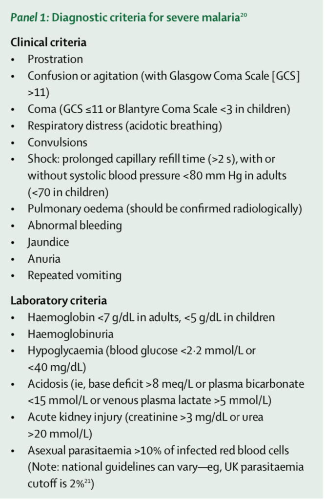
    • Diagnostic criteria for severe malaria:

    Ashley 2018

    • Most common manifestations of severe malaria affect the brain, lungs, and kidneys
      • Patients with cerebral malaria can present encephalopathic or comatose, some severe enough to exhibit extensor posturing, or seizures
      • Can have acute lung injury with a quarter of these patients progressing to ARDS
      • Can have AKI from ATN and resultant acidosis
    • Labs may be unremarkable but watch for anemia and thrombocytopenia
      • Hgb <5 has an OR = 4.9 for death
      • Severe thrombocytopenia has an OR = 2.8
      • Anemia + Thrombocytopenia has an OR = 13.8 (Lampah 2015, PMID 25170106)
    • Watch for hypoglycemia
    • Be mindful of co-infection with salmonella and HIV
      • Obtain BCx, cover with ceftriaxone

    Diagnosis

    • Blood smear
      • Thick smear to increase sensitivity for detecting parasites
      • Thin smear for quantifying parasitemia and species
    • The first smear is positive in over 90% of cases, but if suspicion is high, it has to be repeated BID for 2-3 days for proper exclusion of malaria (CDC 2019)

    Management

    • For uncomplicated, non-severe cases, most patients with falciparum should be admitted, especially those with no prior exposure to malaria parasites
    • Malarone is one of the first line options
      • Check out other suggested regimens from the CDC
    • Important to note that when they take this, ensure they take with milk or food containing fat to enhance absorption
    • Severe Malaria
      • Resuscitative efforts directed at affected organ
      • Can deteriorate rapidly
      • Initiate IV Artesunate if high level of suspicion
        • Requires call to CDC: CDC Malaria Hotline: (770) 488-7788 or (855) 856-4713 (toll-free) Monday–Friday 9am–5pm EST – (770) 488-7100 after hours, weekends, and holidays
      • Benzodiazepines for seizures
      • Be judicious with fluids as this can precipitate pulmonary edema and cerebral edema
        • a/w increased mortality in children at 48 hour
        • (Maitland 2011, PMID: 21615299; Hanson 2013, PMID: 23324951)

    Take Home Points

    • This is going to be a diagnosis that is mainly made through a thorough history, and pay particular attention to those with recent travel to West-Africa
    • The incubation period for falciparum is 12 days, but there is a range of weeks and we should consider Malaria when consistent symptoms develop within 1 month of travel to an endemic area
    • Typical signs and symptoms for uncomplicated malaria are periodic fevers, jaundice, pallor
    • Be mindful of end organ involvement, such as cerebral edema, ATN, and pulmonary edema; these cases are considered to be severe and treated differently than uncomplicated malaria
    • Uncomplicated cases should get Malarone or Coartem
    • Severe cases require IV Artesunate
    • Be judicious with your fluid resuscitation as this can harm our patients

    References

    Centers for Disease Control and Prevention. CDC Parasites – Malaria. 2019 https://www.cdc.gov/parasites/malaria/index.html (7 July 2019, date last accessed)

    Ashley EA, Pyae Phyo A, Woodrow CJ. Malaria. Lancet. 2018;391(10130):1608-21.

    Hanson JP, Lam SW, Mohanty S, Alam S, Pattnaik R, Mahanta KC, et al. Fluid resuscitation of adults with severe falciparum malaria: effects on Acid-base status, renal function, and extravascular lung water. Crit Care Med. 2013;41(4):972-81.

    Lampah DA, Yeo TW, Malloy M, Kenangalem E, Douglas NM, Ronaldo D, et al. Severe malarial thrombocytopenia: a risk factor for mortality in Papua, Indonesia. J Infect Dis. 2015;211(4):623-34.

    Lokken KL, Stull-Lane AR, Poels K, Tsolis RM. Malaria Parasite-Mediated Alteration of Macrophage Function and Increased Iron Availability Predispose to Disseminated Nontyphoidal Salmonella Infection. Infect Immun. 2018;86(9).

    Maitland K, Kiguli S, Opoka RO, Engoru C, Olupot-Olupot P, Akech SO, et al. Mortality after fluid bolus in African children with severe infection. N Engl J Med. 2011;364(26):2483-95.

    Park SE, Pak GD, Aaby P, Adu-Sarkodie Y, Ali M, Aseffa A, et al. The Relationship Between Invasive Nontyphoidal Salmonella Disease, Other Bacterial Bloodstream Infections, and Malaria in Sub-Saharan Africa. Clin Infect Dis. 2016;62 Suppl 1:S23-31.

    Tintanelli, Judith E., et al. Tintinalli’s Emergency Medicine: A Comprehensive Study Guide. Eighth edition. New York: McGraw-Hill Education, 2016: p.1070-1077

    World Health Organization. Guidelines for the treatment of malaria. Third edition

    April 2015. WHO. 2015 https://www.who.int/malaria/publications/atoz/9789241549127/en/ (7 July 2019, date last accessed)

    A special thanks to our editor:

    Angelica Cifuentes Kottkamp, MD
    Infectious Diseases & Immunology
    NYU School of Medicine


    Read More

    Episode 166.0 – Acute Otitis Media Jul 01, 2019
    Show notes

    A look at this common and controversial topic.

    Hosts:
    Brian Gilberti, MD
    Audrey Bree Tse, MD

    https://media.blubrry.com/coreem/content.blubrry.com/coreem/Acute_Otitis_Media.mp3 Download Leave a Comment Tags: Pediatrics

    Show Notes

    Background:

    • The most common infection seen in pediatrics and the most common reason these kids receive antibiotics
    • The release of the PCV (pneumococcal conjugate vaccine), or Prevnar vaccine, has made a big difference since its release in 2000 (Marom 2014)
      • This, along with more stringent criteria for what we are calling AOM, has led to a significant decrease in the number of cases seen since then
      • 29% reduction in AOM caused by all pneumococcal serotypes among children who received PCV7 before 24 months of age
    • The peak incidence is between 6 and 18 months of age
    • Risk factors: winter season, genetic predisposition, day care, low socioeconomic status, males, reduced duration of or no breast feeding, and exposure to tobacco smoke.
    • The predominant organisms: Streptococcus pneumoniae, non-typable Haemophilus influenzae (NTHi), and Moraxella catarrhalis.
      • Prevalence rates of infections due to Streptococcus pneumoniae are declining due to widespread use of the Prevnar vaccine while the proportion of Moraxella and NTHi infection increases with NTHi now the most common causative bacterium
      • Strep pneumo is associated with more severe illness, like worse fevers, otalgia and also increased incidence of complications like mastoiditis.

    Diagnosis

    • The diagnosis of acute otitis media is a clinical one without a gold standard in the ED (tympanocentesis)
    • Ear pain (+LR 3.0-7.3), or in the preverbal child, ear-tugging or rubbing is going to be the most common symptom but far from universally present in children. Parents may also report fevers, excessive crying, decreased activity, and difficulty sleeping.
    • Challenging especially in the younger patient, whose symptoms may be non-specific and exam is difficult
    • Important to keep in mind that otitis media with effusion, which does not require antibiotics, can masquerade as AOM

    AAP: Diagnosis of Acute Otitis Media (2013)*

    1. In 2013, the AAP came out with a paper to help guide the diagnosis of AOM
    2. Moderate-Severe bulging of the tympanic membrane or new-onset otorrhea not due to acute otitis externa (grade B)
      1. The presence of bulging is a specific sign and will help us distinguish between AOM and OME, the latter has opacification of the tympanic membrane or air-fluid level without bulging (Shaikh 2012, with algorithm)
      2. Bulging of the TM is the most important feature and one systematic review found that its presence had an adjusted LR of 51 (Rothman 2003)
      3. Classic triad is bulging along with impaired mobility and redness or cloudiness of TM
    3. Mild bulging of the tympanic membrane AND (grade C)
      1. Recent onset (48hrs)
        Ear pain (verbal child)
        Holding, tugging, rubbing of the ear (non-verbal child)
        OR
      2. Intense erythema of the tympanic membrane

    * The diagnosis should not be made in the absence of a middle ear effusion (grade B)

    Treatment Options

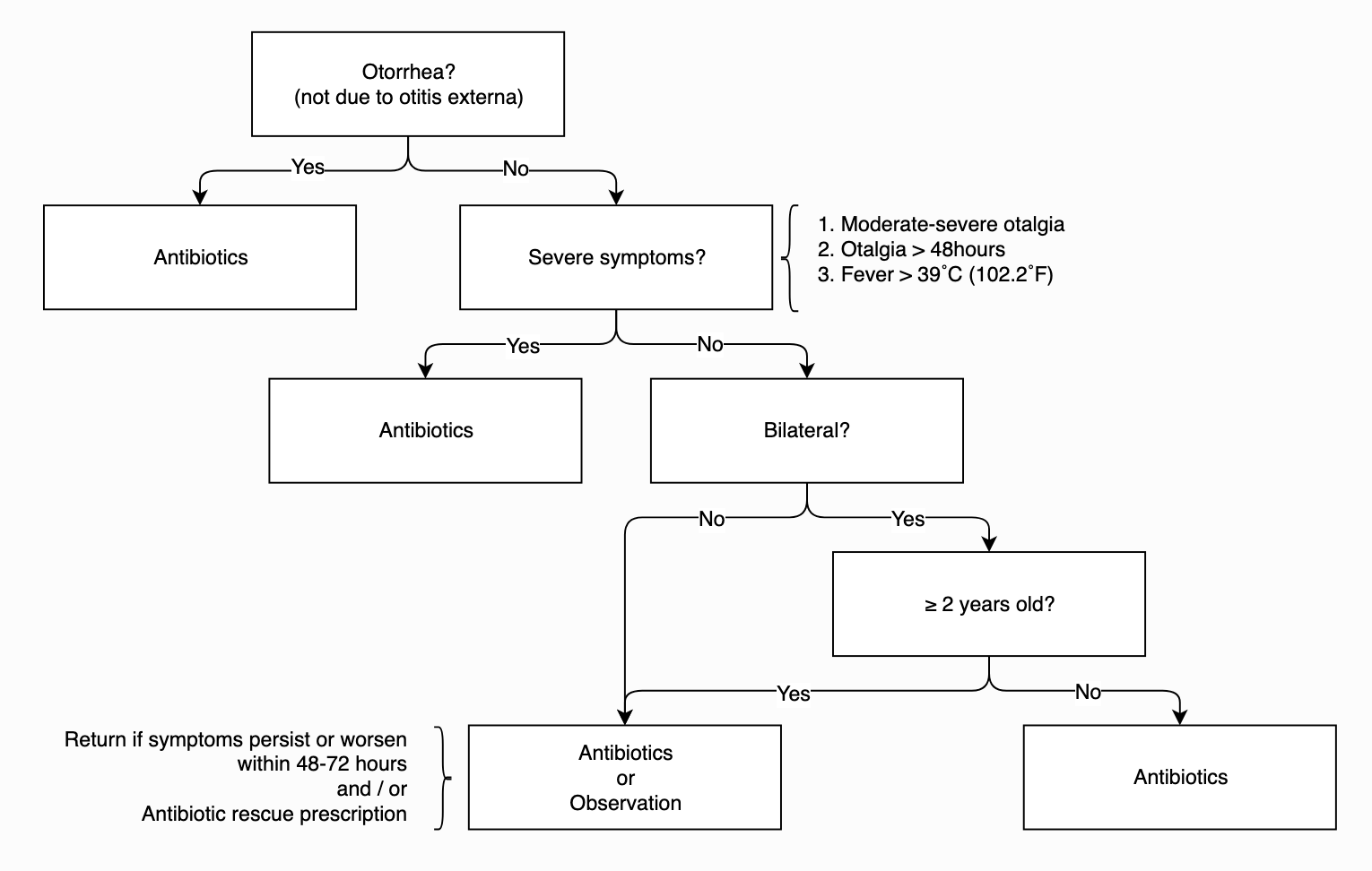
    • A strategy of “watchful waiting” in which children with acute otitis media are not immediately treated with antibiotic therapy, has been endorsed by the American Academy of Pediatrics.
    • Who gets antibiotics?
      • Depends on age, temperature, duration of otalgia, laterality / otorrhea, and access to follow up
      • Get’s antibiotics:
        • <6 months: Treat
        • 6 months to 2 years: Treat
          • Exception, AAP permits initial observation: unilateral AOM with mild symptoms (mild ear pain, <48h, T <102.2)
          • But know that there is a high rate of treatment failure (Hoberman 2013)
        • >2: Treat
          • Unless they have mild symptoms and it’s unilateral, you can observe for 48-72 hours
        • Why do we give antibiotics?
          • Demonstrated reduction in pain, TM perforations, contralateral episodes of AOM
          • They are no walk in the park, with increased adverse events (vomiting, diarrhea, rash)
        • Two well-designed clinical trials (2011) randomized approximately 600 children meeting strict diagnostic criteria for acute otitis media to receive Augmentin or placebo. These studies demonstrated a significant reduction in symptom burden and clinical failures in those who received antibiotics.
        • The authors conclude that those patients with a clear diagnosis of acute otitis media would benefit from antibiotic therapy

    AAP AOM Treatment Algorithm
    Antibiotic Selection

    • High-dose amoxicillin in most (for now)
    • Amoxicillin should not be used if the patient has received Amoxicillin in the past 30 days, has concomitant purulent conjunctivitis (likely H flu) or is allergic to penicillin.
      • beta lactamase resistant antibiotic should be used.
        • Amoxicillin clavulanate or 2nd or 3rd generation cephalosporins (including intramuscular ceftriaxone).
      • Patients with a history of type 1 hypersensitivity reactions to penicillin should be treated macrolides.
      • Studies on duration of therapy have shown better results with 10-day duration in children younger than 2 years and suggest improved efficacy in those 2-5 years.
      • For patients older than 5 years, shorter course therapy (5-7 days) can be utilized.

    Pain Control

    • Motrin and APAP may have benefit with otalgia reduction

    Other

    • Decongestants and antihistamines have been shown to not benefit patients in terms of duration of symptoms or complication rate. Not surprisingly, these agents increase the side-effects experienced by patients.

    Follow up

    • If you chose to observe, let the parents know to return to ED or f/u with their provider in 48-72 hours if they symptoms do not improve. Providing a prescription to parents with clear instructions on when to fill it is also an acceptable option. Strict return precautions should be given if patient develops meningismus or facial nerve palsy.
    • If antibiotics were initiated, and there isn’t improvement in 2-3 days, the diagnosis of AOM should be revisited and, if still suspected, we have to consider that the causative bug is resistant to the prescribed antibiotic.
      • These patients should RTED or f/u with their pediatrician for escalation of care
        • Amoxicillin → Augmentin
        • Augmentin → Ceftriaxone IM
        • Macrolide → no clear antimicrobial agent, consult pediatric ENT
      • If antibiotics are initiated with resolution of symptoms, the patient should f/u in 2-3 months to ensure resolution of the middle ear effusion and ensure that there is no associated conductive hearing loss

    References:

    Coker TR, Chan LS, Newberry SJ, Limbos MA, Suttorp MJ, Shekelle PG, et al. Diagnosis, microbial epidemiology, and antibiotic treatment of acute otitis media in children: a systematic review. JAMA. 2010;304(19):2161-9.

    Hoberman A, Ruohola A, Shaikh N, Tahtinen PA, Paradise JL. Acute otitis media in children younger than 2 years. JAMA Pediatr. 2013;167(12):1171-2.

    Lieberthal AS, Carroll AE, Chonmaitree T, Ganiats TG, Hoberman A, Jackson MA, et al. The diagnosis and management of acute otitis media. Pediatrics. 2013;131(3):e964-99.

    Marom T, Tan A, Wilkinson GS, Pierson KS, Freeman JL, Chonmaitree T. Trends in otitis media-related health care use in the United States, 2001-2011. JAMA Pediatr. 2014;168(1):68-75.

    Rothman R, Owens T, Simel DL. Does this child have acute otitis media? JAMA. 2003;290(12):1633-40.

    Shaikh N, Hoberman A, Rockette HE, Kurs-Lasky M. Development of an algorithm for the diagnosis of otitis media. Acad Pediatr. 2012;12(3):214-8.

    Venekamp RP, Sanders S, Glasziou PP, Del Mar CB, Rovers MM. Antibiotics for acute otitis media in children. Cochrane Database Syst Rev. 2013(1):CD000219.

    See our core article on the topic by Dr. Deborah Levine and Dr. Michael Mojica here

    A special thanks to our editors:

    Michael A. Mojica, MD

    Director, Pediatric Emergency Medicine Fellowship
    Bellevue Hospital Center

    Christie M. Gutierrez, MD

    Pediatric Emergency Medicine Fellow
    Columbia University Medical Center
    Morgan Stanley Children’s Hospital
    New York Presbyterian


    Read More

    Episode 165.0 – Foot Fractures Jun 17, 2019
    Show notes

    A look at foot fractures – which can be splinted and which may need the OR.

    Hosts:
    Audrey Bree Tse, MD
    Brian Gilberti, MD

    https://media.blubrry.com/coreem/content.blubrry.com/coreem/Foot_Fractures.mp3 Download Leave a Comment Tags: Orthopedics

    Show Notes

    Episode Produced by Audrey Bree Tse, MD

    Background:

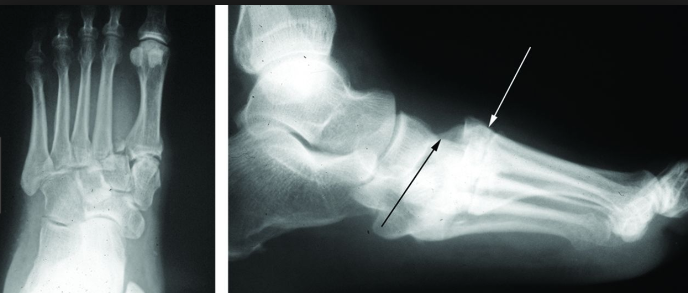
    • Why do we care about Jones fractures?
      • Propensity for poor healing due to watershed area of blood supply
    • Fifth metatarsal fractures account for 68% of metatarsal fractures in adults
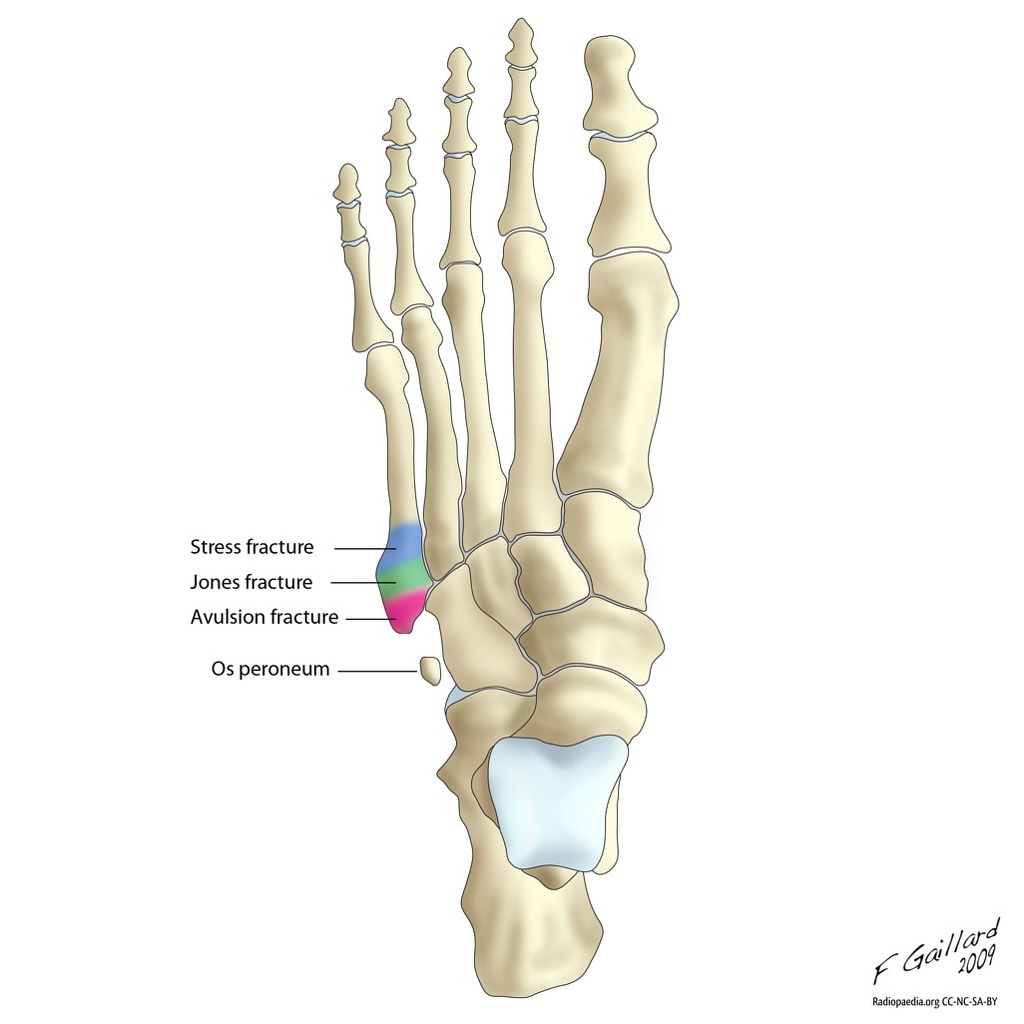
    • Proximal 5th metatarsal fractures are divided into 3 zones (93% zone 1, 4% zone 2, 3% zone 3)
    • Zone 1 (pseudo-Jones):
      • Tuberosity avulsion fracture
      • Typically avulsion type injuries due to acute episode of forefoot supination with plantar flexion
      • Typical fracture pattern is transverse to slightly oblique
    • Zone 2 (Jones fracture):
      • Fracture at the metaphyseal-diaphyseal junction of the fifth metatarsal
      • Typically acute episode of large adduction force applied to forefoot with the ankle plantar flexed
    • Zone 3:
      • Proximal diaphyseal stress fracture
      • Typically results from a fatigue or stress mechanism

    Clinical Presentation:

    • History of acute or repetitive trauma to forefoot
    • Fracture type / pattern closely related to injury location
    • Foot often swollen, ecchymotic, very tender to fifth metatarsal +/- crepitus, inability to hear weight

    Diagnosis:

    • Clinical exam:
      • Evaluate skin integrity
      • Check neurovascular status
      • Evaluate toes/ feet/ ankles/ tib fib/ knees/ hips, involved tendon function, associated adjacent structures (Achilles, ankle ROM/ function, etc)
    • 3 XR views: lateral, anteroposterior, 45* oblique
    • Acute stress fractures are typically not detected on the standard 3 views; therefore, repeat XRs 10-14d after onset of sx (may see radiolucent reabsorption gap around fracture)
    • For more complex mid foot trauma, consider CT to r/o Lisfranc

    Treatment:

    • Consider classification of fracture, patient demographics & activity level when deciding on treatment
    • Tertiary care centers that have access to Orthopedics/Podiatry services
      • Consider consultation for “true” Jones fractures, as some cases may be operatively managed acutely and/or for expedited follow-up to be arranged
    • If working in community/rural locations: other than patients that present with “open” injuries, concerns for compartment syndrome (almost never), and “high-end”/professional athletes, there are generally no other circumstances that would require expedited transfer to a tertiary care center for immediate further evaluation.
    • Less favorable outcomes associated with certain patient factors: female gender, DM, obesity

    Surgical:

    • Different modalities of surgery:
      • Intramedullary screw
      • Bone graft
      • Closed reduction and fixation with K-wire
      • ORIF (all +/- need for bone graft)
    • Surgery likely recommended for displacement >10 degrees of plantar angulation or 3-4 mm of translation in any plane
    • Indications for OR:
      • Neck and shaft fractures with >10 degrees plantar angulation or 3mm of displacement in any plane with insufficient closed reduction
      • Avulsion fractures (zone one) with >3 mm of displacement or comminuted
      • Zone two fractures: displaced zone two fractures require operative management. For acute non displaced Jones fractures, consider early intramedullary screw fixation in athletes (studies have shown return to sport ~ 8 weeks, weight bearing within 1-2 weeks)
      • Zone three fractures (diaphyseal stress fractures) in athletes

    Nonoperative:

    • All non displaced fifth metatarsal fractures can be treated non operatively
    • Non displaced zone 1 fractures: protected weight bearing/ symptomatic care in short leg walking cast, air-boot, posterior splint, or compression wrap/ rigid shoe until discomfort subsides
    • Zone 2 and 3 fractures are more complex because they often result in prolonged healing time and potential for delayed/ nonunion
    • Acute zone 2 fractures: nonweightbearing in short leg cast for 6-8 weeks
    • Acute zone 3 fractures: nonweightbearing in short leg cast for up to 20 weeks
    • With respect to athletes: repeat fracture after surgical treatment of Jones fracture can occur after healing and screw removal; thus it is recommended that the screw be left in until the end of the athlete’s career

    References:

    Bowes J, Buckley R. Fifth metatarsal fractures and current treatment. World J Orthop. 2016;7(12):793–800. Published 2016 Dec 18. doi:10.5312/wjo.v7.i12.793

    Petrisor BA, Ekrol I, Court-Brown C. The epidemiology of metatarsal fractures. Foot Ankle Int. 2006 Mar; 27(3): 172-4.

    Rammelt S, Heineck J, Zwipp H. Metatarsal fractures. Injury. 2004;35 Suppl 2:SB77–SB86.

    Tham W, Sng S, Lum YM, Chee YH. A Look Back in Time: Sir Robert Jones, ‘Father of Modern Orthopaedics’. Malays Orthop J. 2014;8(3):37–41. doi:10.5704/MOJ.1411.009

    Thomas JL, Davis BC. Three-wire fixation technique for displaced fifth metatarsal base fractures. J Foot Ankle Surg. 2011;50:776–779.

    ______________________

    LISFRANC SHOW NOTES:

    Intro:

    • Can’t miss diagnoses: needs stat ortho
    • 20% miss rate
    • Can be dislocation, fracture, fracture dislocation, or ligamentous injury
    • Jacques Lisfranc in Napoleonic Wars: performed transmetatarsal amputation for midfoot gangrene

    Anatomy:

    • Lisfranc ligament: 3 ligaments that run from the base of the second metatarsal to the medial cuneiform bone. Helps attach the forefoot to the midfoot bones
    • If ligament complex gets disrupted, can end up with chronic deformity and disability

    Injury definitions:

    • Dislocation: widening between base of 1st and 2nd metatarsal, or between cuneiforms
    • Fracture dislocation: associated fracture, most commonly at the base of the proximal second metatarsal

    Physical Exam:

    • Pain and swelling in midfoot
    • Pain elicited with passive abduction and pronation of the midfoot while holding heel steady
    • Plantar ecchymosis
    • r/o compartment syndrome
    • Feel for DP pulse!

    Diagnosis:

    • XRs: AP, lateral, oblique, stress views with weight bearing
    • Watch out for “fleck sign”
    • Consider CT if pt cannot bear weight, or even if XR negative and high suspicion

    Treatment:

    • Ortho consult!

    ______________________

    THANKS TO DANNY PURCELL, MD and MAY LI, MD


    Read More

    Episode 164.0 – Debriefing Jun 03, 2019
    Show notes

    A discussion with Drs. McNamara and Leifer on the essentials and beyond of debriefing

    Hosts:
    Brian Gilberti, MD
    Audrey Tse, MD

    https://media.blubrry.com/coreem/content.blubrry.com/coreem/Debriefing.mp3 Download One Comment Tags: Resuscitation, Simulation

    Show Notes

    TAKE HOME POINTS

    • Debriefing after a clinical case in the ED is a way to have an interprofessional, reflective conversation with a focus on improving for the next patient. We can debrief routine cases, challenging cases, or even cases that go well.
    • Follow a structure when leading a debrief.
      • The prebrief sets ground rules and informs the team that the debrief is optional and will only take 3-5 minutes.
      • Introduce names and roles
      • Then give a one-liner about what happened in the case, followed by a plus/ delta: address what went well and why, then how to improve
      • Finally, wrap up with take home points
    • Pitfalls to watch out for in clinical debriefing include:
      • Avoid siloing or alienating any learners. Learn from all your colleagues on your team- it’s less about medicine and more about interprofessional and systems issues
      • Don’t pick on individual performance. It’s not about shaming- it’s about improving patient care
      • Avoid “guess what I’m thinking” questions; ask real questions
      • Proceed with caution in order to dampen or avoid psychological trauma and second victim syndrome. The learner may ask “was this my fault?”; we never want a learner to feel this way. Ask, what systems supported or did not support you today? Talk about what happened. Avoid shame and blame.
    • Have the right values and do it for the right reasons.

    ADDITIONAL TOOLS

    PEARLS Debriefing Tool

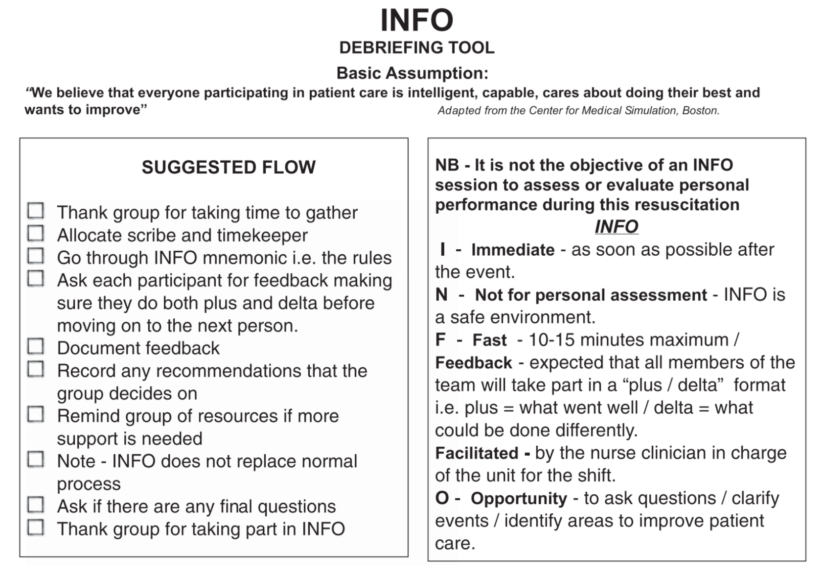
    INFO Model:

    GUESTS

    Dr. Shannon McNamara completed residency in Emergency Medicine at Temple University hospital and fellowship in Medical Simulation at Mount Sinai St. Lukes-Roosevelt. She now is the Director of the Simulation Division in the NYU Department of Emergency Medicine. She’s thrilled to have somehow made a career out of teaching people to talk about their feelings using big computers shaped like people.

    Dr. Jessica Leifer attended NYU for medical school and completed her residency training in emergency medicine at Mount Sinai St. Luke’s-Roosevelt. She completed a fellowship in medical simulation at the Mount Sinai Hospital. She is now simulation faculty in the NYU department of Emergency Medicine. Her academic interests include using simulation for patient safety, operations, and improving teamwork.


    Read More

    Episode 163.0 – Croup May 20, 2019
    Show notes

    A look at one of the most common and potentially concerning upper respiratory infections in children.

    Host:
    Brian Gilberti, MD

    https://media.blubrry.com/coreem/content.blubrry.com/coreem/Croup.mp3 Download One Comment Tags: Airway, Infectious Diseases, Pediatrics

    Show Notes

    Background

    • Croup is a viral infection starts in the nasal and pharyngeal mucosa but spreads to the larynx and trachea
      • Subglottic narrowing from inflammation
      • Dynamic obstruction
      • Barking cough
      • Inspiratory stridor
    • Causes:
      • Parainfluenza virus (most common)
      • Rhinovirus
      • Enterovirus
      • RSV
      • Rarely: Influenza, Measles
    • Age range: 6 months to 36 months
    • Seasonal component with high prevalence in fall and early winter
    • Differential
      • Bacterial tracheitis
      • Acute epiglottitis
      • Inhaled FB
      • Retropharyngeal abscess
      • Anaphylaxis

    Presentation & Diagnosis

    • Classically a prodrome of nonspecific symptoms for 1-3 days with low grade fevers, congestion, runny nose.
    • Symptoms reach peak severity on the 4th day
    • “Steeple sign” on Xray (subglottic narrowing) present in only 50% of patients with croup
    • Assess air entry, skin color, level of consciousness, for tachypnea, if there are retractions / nasal flaring (if present at rest or with agitation) & coughing
    • “Westley Croup Score” (https://www.mdcalc.com/westley-croup-score)
      • Chest wall retractions
      • Stridor
      • Cyanosis
      • Level of consciousness
      • Air entry

    Management

    • Mild Croup
      • Occasional barking cough, but no stridor at rest and mild to no retractions
      • Tx: Single dose of dex
        • Has been shown to improve severity and duration of symptoms
        • Route is not particularly important, whether it’s PO, IV or IM
        • Chosen route should aim to minimize agitation in the patient that might worsen their condition
      • May be managed at with supportive care
        • Humidifiers (NB: there isn’t good evidence supporting the use of humidifiers)
        • Antipyretics
        • PO fluids
      • Moderate Group
        • May have stridor at rest, mild-moderate retractions but no AMS and will not be in distress.
        • Tx: Dex + Racemic Epinephrine
          • Racemic epinpehrine will start to work in about 10 minutes
          • Effects last for more than an hour
        • Severe group
          • Receives the same initial therapy as the moderate group with dex and race epi
          • Pts with worrisome signs: stridor at rest, marked retraction, cyanosis and/or lethargy
          • Heliox (a combinations of 70-80% helium + 20-30% oxygen) may be attempted
            • There is limited evidence to support the role of heliox in croup,
            • NB: Pt may require higher levels of oxygen than the 20-30% mixture may provide
          • Intubation
            • Anticipate edema narrowing the airway
            • Consider starting with a tube that is 0.5 to 1 mm smaller than size typically used

    Disposition:

    • Patients without stridor at rest or respiratory distress can be generally discharged from the ED
    • If epinephrine is given, patients should be monitored for 2-4 hours for reemergence of symptoms as the medication wears off

    Take Home Points

    • Croup usually affects children within the age range of 6 months to 36 months with the most common cause being parainfluenza virus
    • Given the symptom overlap, we must consider more concerning diagnoses, including bacterial tracheitis, in these patients, especially if they are ill appearing or traditional therapies are ineffective
    • All patients benefit from a one-time dose of dexamethasone and, if racemic epinephrine is given, the patient should be observed for at least 3 hours
    • If intubation is required, anticipate a narrowed airway

    Parent Article: https://coreem.net/core/croup/ by Dr. Pankow


    Read More

    Episode 162.0 – Stevens-Johnson Syndrome/Toxic Epidermal Necrolysis May 06, 2019
    Show notes

    A look at this deadly mucocutaneous reaction and how to best manage these patients in the ED

    https://media.blubrry.com/coreem/content.blubrry.com/coreem/SJS.mp3 Download Leave a Comment Tags: Critical Care, Dermatology

    Show Notes

    Episode Produced by Audrey Bree Tse, MD

    • Rash with dysuria should raise concern for SJS with associated urethritis
      • Dysuria present in a majority of cases
    • SJS is a mucocutaneous reaction caused by Type IV hypersensitivity
      • Cytotoxic t-lymphocytes apoptose keratinocytes → blistering, bullae formation, and sloughing of the detached skin
    • Disease spectrum
      • SJS = <10% TBSA
      • TEN = >30% TBSA
      • SJS/ TEN Overlap = 10-30% TBSA
    • Incidence is estimated at around 9 per 1 million people in the US
    • Mortality is 10% for SJS and 30-50% for TEN
      • Mainly 2/2 sepsis and end organ dysfunction.
    • SJS can occur even without a precipitating medication
      • Infection can set it off especially in patients with risk factors including HIV, lupus, underlying malignancy, and genetic factors
      • SATAN for the most common drugs
        • Sulfa, Allopurinol, Tetracyclines, Anticonvulsants, and NSAIDS
      • Anti-epileptics include carbamazepine, lamictal, phenobarb, and phenytoin
    • Can have a curious course
      • Hypersensitivity reaction can develop while taking medication, or even one to four weeks after exposure
      • In pediatric population, mycoplasma pneumonia and herpes simplex have been identified as precipitating infections
    • Patients often have a prodrome 1-3 days prior to the skin lesions appearing
      • May complain of fever, myalgias, headaches, URI symptoms, and malaise
    • Rash may be the sole complaint
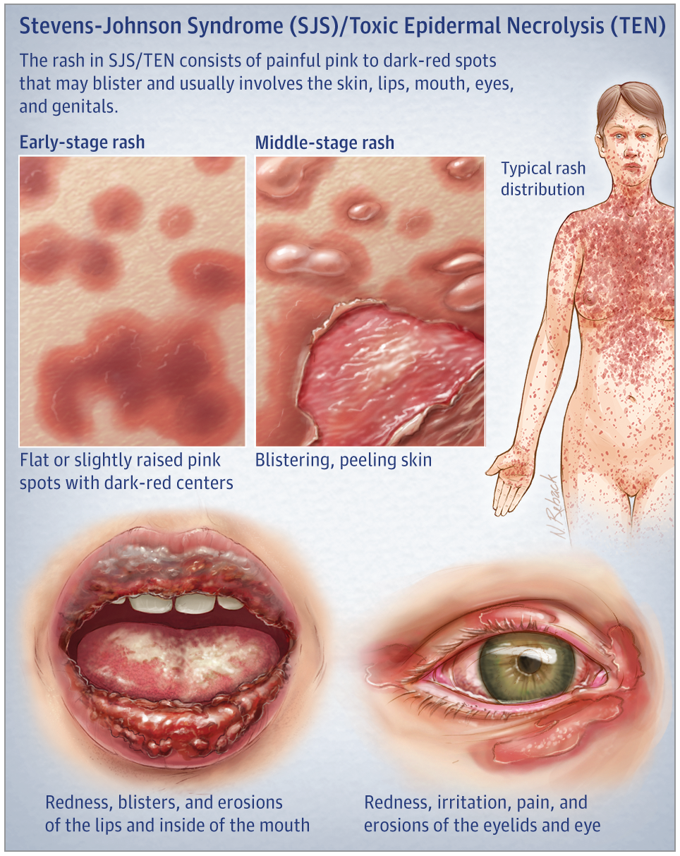
      • Starts as dark purple or erythematous lesions with purpuric centers that progress to bullae
      • Skin surrounding the lesions detaches from the dermis with just light pressure (Nikolsky Sign)
      • Up to 95% of patients will have mucous membrane lesions
      • ~85% will have conjunctival lesions
      • Symptoms: Burning or itching eyes, a cough or sore throat, pain with eating, pain with urinating or defecating

    Source: JAMA Dermatol. 2017

    • Differential Diagnosis: SSSS, autoimmune bullous diseases, bullous fixed drug eruption, erythema multiforme, thermal burns, phototoxic reactions, and TSS
    • SJS is a clinical diagnosis
      • Basic workup: CBC, chemistry panel, LFTs, and a UA
    • Treatment
      • Supportive care
        • IV fluid repletion guided by TBSA affected, as well as electrolyte, protein, and energy supplementation
      • Consider protecting airway if significant oral mucosal involvement
      • Stop the offending agent (if there is one)
      • Advanced wound care and pain control
      • Consults: Derm to do a biopsy, +/- ophthalmology, gyn / urology to prevent strictures or contractures
      • Consider transferring to a burn center
    • Dispo:
      • Low threshold for ICU admission
      • SCORTEN ( max of 7 points)
        • 1 point each for
          • Age over 40
          • Current cancer
          • >30% body surface area affected
          • HR >120
          • BUN >28
          • Glucose >240
          • Bicarb <20
          • Score of 2 points or higher should -> ICU

    Take Home Points

    • SJS may begin like the flu, with lesions appearing 1-3 days after the prodrome starts
    • Have to have a high suspicion for SJS because it is deadly. It’s a clinical diagnosis — derm biopsy is supportive
    • A thorough history and physical exam are key. Remember the characteristic rash and bullae, and always look in the mouth and eyes. Ask about dysuria, sore throat, and eye irritation, as well as preceding medications or infections. Think SATAN!
    • Prompt supportive care focused on ABCs and IVF repletion are critical. These patients can get sick really fast, so consider an ICU or burn unit.

    References:

    Barrett W. Quick Consult: Symptoms: Rash, Dysuria, and Mouth Sores. Emergency Medicine News. 41(4): 15-16, April 2019.

    Bivins H, Comes J. Stevens-Johnson Syndrome. Rosen and Barkin’s 5-Minute Emergency Medicine Consult. 2015; 1076-1077.

    Ergen EN, Hughey LC. Stevens-Johnson Syndrome and Toxic Epidermal Necrolysis. JAMA Dermatol.2017;153(12):1344. doi:10.1001/jamadermatol.2017.3957

    Gerull R, Nelle M, Schaible T. Toxic epidermal necrolysis and Stevens-Johnson syndrome: A review. Crit Care Med. 2011; 39:1521-1532.

    Lerch M, Mainetti C, Terziroli Beretta-Piccoli B, Harr T. Current Perspectives on Stevens-Johnson Syndrome and Toxic Epidermal Necrolysis. Clin Rev Allergy Immunol. 2018;54(1):147-76.

    McNeil, D. (2019). Measles Cases Surpass 700 as Outbreak Continues Unabated. [online] Nytimes.com. Available at: https://www.nytimes.com/2019/04/29/health/measles-outbreak-cdc.html [Accessed 6 May 2019].

    Mustafa SS, Ostrov D, Yerly D. Severe Cutaneous Adverse Drug Reactions: Presentation, Risk Factors, and Management. Curr Allergy Asthma Rep. 2018;18(4):26.


    Read More

    Episode 161.0 – Opioid Epidemic Apr 22, 2019
    Show notes

    A look at the opioid epidemic and what ED providers can do to combat this formidable foe.

    https://media.blubrry.com/coreem/content.blubrry.com/coreem/Opioid_Epidemic.mp3 Download Leave a Comment Tags: Opioid Dependence, Opioid Free ED

    Show Notes

    • Consider alternatives to opiates for acute pain
      • NSAIDs
      • Subdissociative ketamine
      • Nerve blocks
    • Curb misuse and diversion through prescribing a short supply and perform I-STOP checks
    • Narcan is not just for acute overdose treatment by EMS or within the ED anymore
      • We can equip patients, family members and friends with Narcan kits prior to discharge
      • In New York state, can prescribe Narcan to patients with near fatal overdoses or who screen positive for an opioid use disorder
      • Intranasal formulation is cheaper and more commonly prescribed than IM
    • Buprenorphine induction can be done in the ED for patients in active withdrawal, as calculated by the COWS score.
      • MDcalc calculator: https://www.mdcalc.com/cows-score-opiate-withdrawal
      • Providers do not need an X-waiver to give a dose of Buprenorphine in the ED for 3 days
    • Home induction can be considered for patients not actively withdrawing but would like to enter medication assisted treatment
      • Some considerations:
        • Contraindicated in patients with severe liver dysfunction and with hypersensitivity reaction to drug
        • Oversedation can occur with concurrent use of benzodiazepines and alcohol
        • Will precipitate withdrawal if concurrently using full opioid agonists
      • Longitudinal care has to be established for patients started on Buprenorphine
        • SAMHSA’s Buprenorphine practitioner locator site: https://www.samhsa.gov/medication-assisted-treatment/practitioner-program-data/treatment-practitioner-locator
        • Buprenorphine Induction Pamphlet

    Read More

    Episode 160.0 – Measles Apr 08, 2019
    Show notes

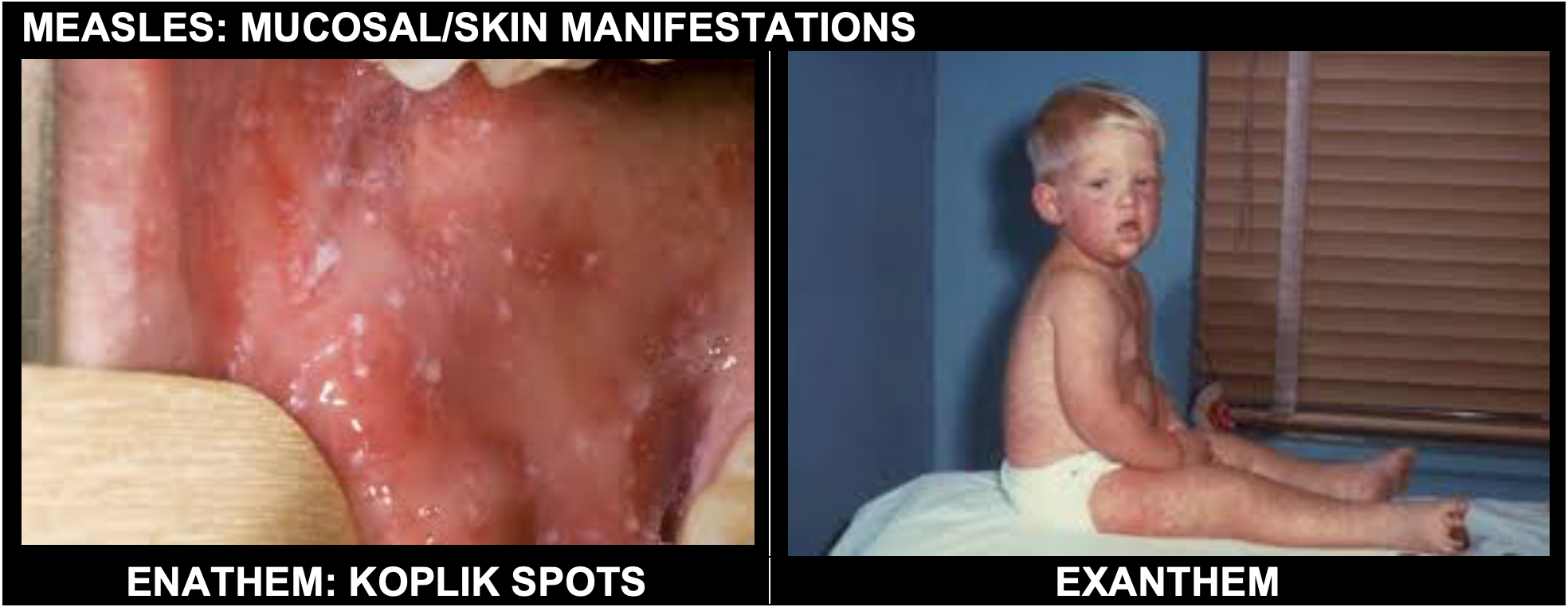
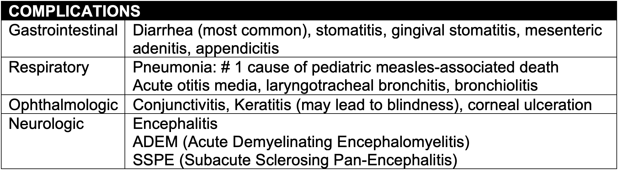
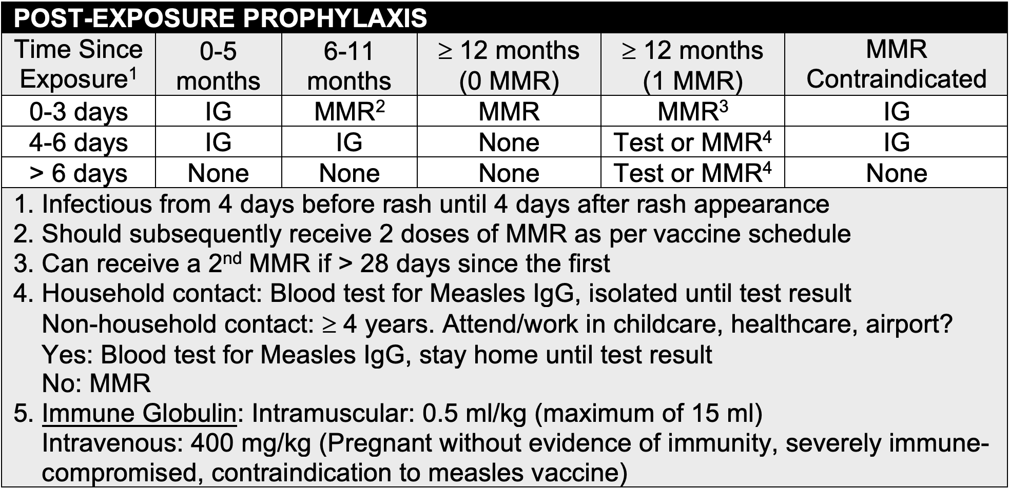
    In this episode, we discuss the recent measles outbreak and how ED providers can best prepare to treat this almost vanquished foe.

    https://media.blubrry.com/coreem/content.blubrry.com/coreem/Measles_Final_Cut.mp3 Download One Comment Tags: Infectious Diseases, Pediatrics

    Show Notes

    Episode Produced by Audrey Bree Tse, MD

    References:

    CDC Measles for Health Care Providers. https://www.cdc.gov/measles/hcp/index.html#lab.

    Gladwin M, Trattler B. Orthomyxo and Paramyxoviridae. In: Clinical Microbiology Made Ridiculously Simple. 4th ed. Miami, FL: MedMaster, Inc; 2009: 240-243.

    Hussey G, Klein M. A Randomized, Controlled Trial of Vitamin A in Children with Severe Measles. N Engl J Med. 1990; 323: 160-164.doi: 10.1056/NEJM199007193230304.

    Nir, Sarah Mailin and Gold, Michael. “An Outbreak Spreads Fear: Of Measles, of Ultra-Orthodox Jews, of Anti-Semitism.” New York Times [New York City] 03/29/2019. https://www.nytimes.com/2019/03/29/nyregion/measles-jewish-community.html

    A massive thanks to:

    Shweta Iyer, MD: NYU Langone 3rd year Pediatric Emergency Medicine Fellow.

    Jennifer Lighter, MD: Assistant Professor of Pediatric Infectious Diseases, NYU School of Medicine.

    Michael Mojica, MD: Associate Professor of Pediatric Emergency Medicine, NYU Langone Medical Center.

    Michael Phillips, MD: Chief Hospital Epidemiologist, NYU Langone Medical Center.


    Read More

    Episode 159.0 – Acute Decompensated Heart Failure Mar 22, 2019
    Show notes

    In this episode, we discuss acute decompensated heart failure and how to best manage these dyspneic patients in the ED.

    https://media.blubrry.com/coreem/content.blubrry.com/coreem/Podcast_ADHF.mp3 Download Leave a Comment Tags: Cardiology, Respiratory

    Show Notes

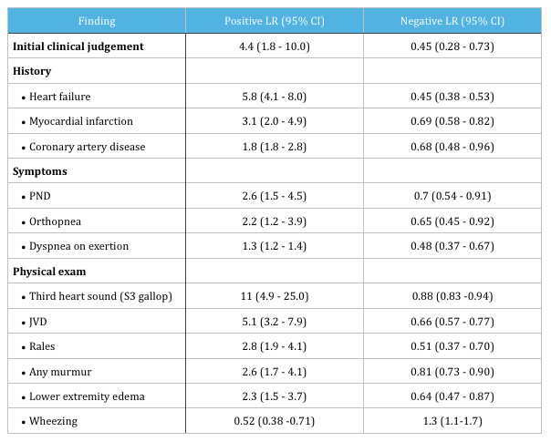
    • Features that increase the probability of heart failure. (Wang 2005)

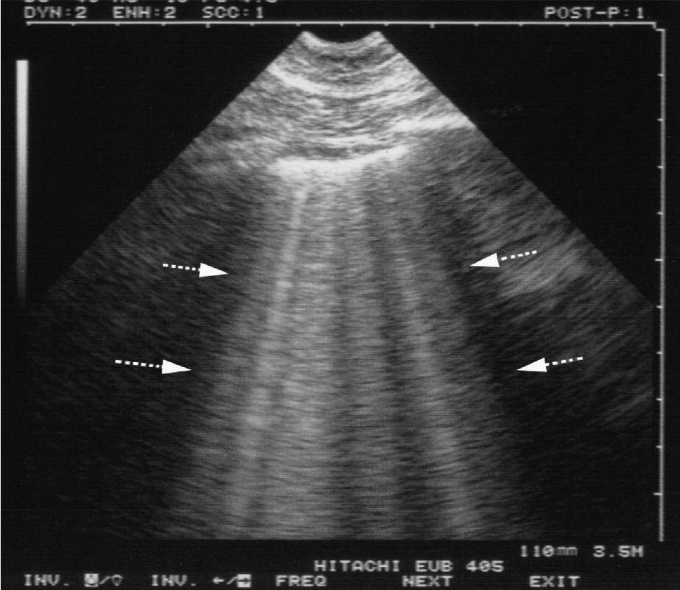
    • B-lines seen in pulmonary edema.

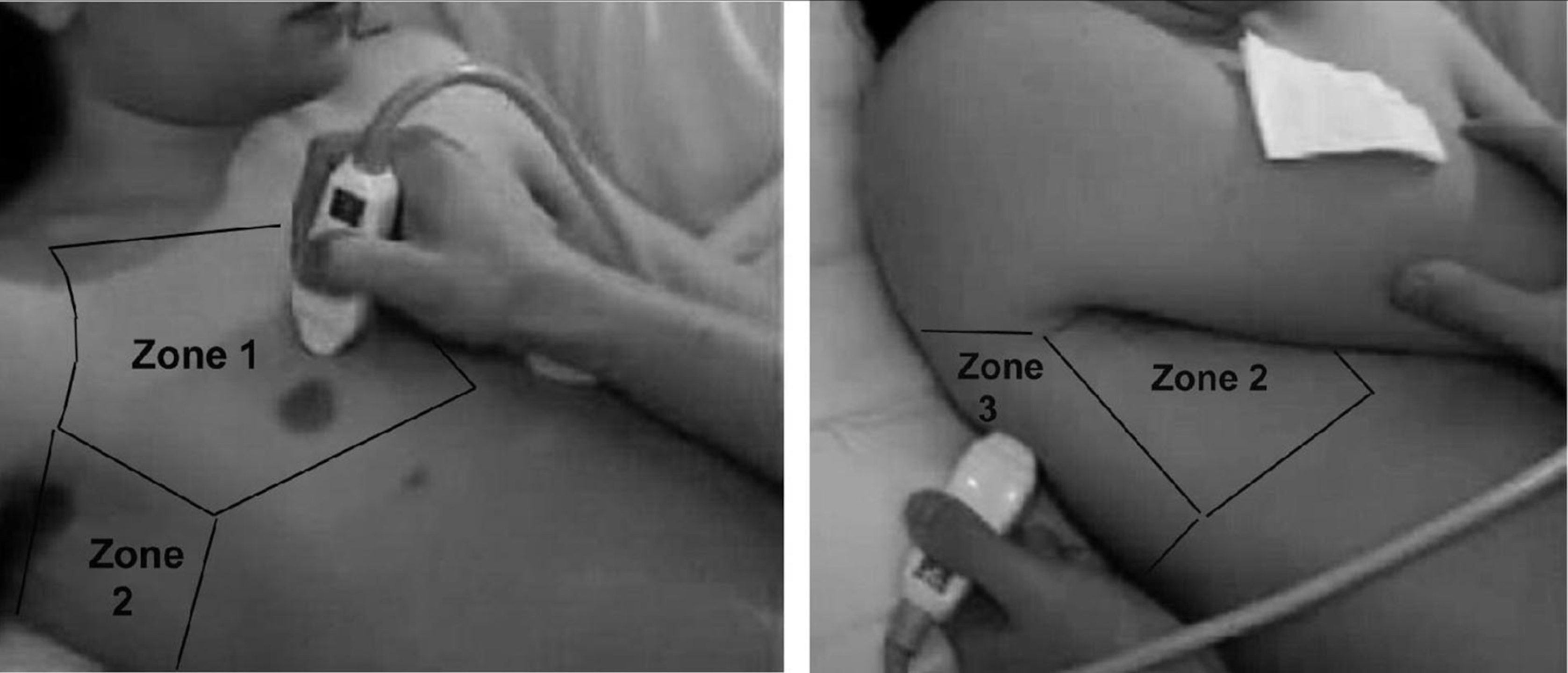
    • Positioning of ultrasound probe in BLUE protocol. (Lichtenstein 2008)


    Read More

    Episode 158.0 – Boxer’s Fracture Mar 08, 2019
    Show notes

    In this episode, we discuss Boxer's fractures and how to best manage them in the ED.

    https://media.blubrry.com/coreem/content.blubrry.com/coreem/Boxer_s_Fracture_eq.m4a Download One Comment Tags: Orthopedics, Trauma

    Podcast Video

    https://youtu.be/UreET5eLHas

    Show Notes

    Background:

    • 40% of all hand fractures
    • A metacarpal fracture can occur at any point along the bone (head, neck, shaft, or base)
    • “Boxer’s” fractures classically at neck
    • Most common mechanism: direct axial load with a clenched fist
    • Most common metacarpal injured is the 5th
    • A majority of these injuries are isolated injuries, closed and stable

    Examination:

    • Ensure that this is an isolated injury
    • May note a loss of knuckle contour or shortening
    • A thorough evaluation of the skin is important
      • Patients may also have fight bites and require irrigation and antibiotics
    • Tender along the dorsum of the affected metacarpal
    • Evaluate the range of motion as the commonly seen shortening results in extension lag
      • For every 2 mm of shortening there is going to be a 7 degree decrease in ability to extend the joint
    • Check rotational alignment of digits with the MCP and PIP at 50% flexion.
      • Partially clench their fist and ensure that the axis of each digit converges near the scaphoid pole / mid wrist
    • Deformity is often seen due to the imbalance of volar and dorsal forces
      • Dorsal angulation
    • AP, lateral and oblique views should be obtained on XR
    • The degree of angulation is estimated with the lateral view
      • NB: Normal angle between the metacarpal head and neck is 15 degrees

    Management:

    • Most may be splinted with an ulnar gutter splint
      • Must be closed, not significantly angulated, and not malrotated
    • When splinting, place the wrist in slight extension, MCP (knuckles) at 90 degrees and the DIP and PIP in a relaxed, slightly flexed position
    • A closed reduction is indicated if there is significant angulation
      • “20, 30, 40” rule
        • If angulation is more than:
          • 20 in the middle finger metacarpal
          • 30 in the ring finger metacarpal
          • 40 in the pinky finger metacarpal
        • Analgesia with a hematoma block or ulnar nerve block
        • Reduction technique: https://www.aliem.com/2013/01/trick-of-trade-reducing-metacarpal/

    Referral:

    • May have mild deformity or decreased functionality and strength in hand grip after this injury
    • Emergent evaluation if:
      • Open fracture
      • Neurovascular compromise

    Follow up:

    • Refer to hand specialist
      • Within 1 week if fractures of 4thand 5thmetacarpals with angulation
      • 3 to 5 days if the 2ndand 3rd metacarpalsare affected
      • Immobilized for three to four weeks in splint
      • Healing may take up to six weeks

    Take Home Points:

    • This is one of the most common fractures we will see as emergency physicians
    • When evaluating these patients, ensure that this are no other more severe, life-threatening injuries, and pay particular attention to the skin exam so that you do not miss a fight-bite
    • Reductions may be required if there is significant angulation, which is guided by the 20, 30, 40 rule
    • Finally, emergent specialist evaluation is indicated if there is an open fracture or evidence of neurovascular compromise

    Read More

    Previous 1 5 6 7 8 9 24 Next

    Related Podcasts

    The Joe Rogan Experience

    1

    The Joe Rogan Experience Comedy
    More Perfect

    2

    More Perfect Government
    Casefile True Crime

    3

    Casefile True Crime Games & Hobbies
    Reply All

    4

    Reply All Games & Hobbies
    You Must Remember This

    5

    You Must Remember This Games & Hobbies
    Real Time with Bill Maher

    6

    Real Time with Bill Maher Games & Hobbies
    footer-logo

    Contact Us

    Toll Free: 844-670-7747

    Links

    • Home
    • Top Charts
    • Networks
    • Apps
    • Independents Podcasts
    • Podcast Advertising
    • Podcast News
    • Contact Us
    • About Us
    • Analytics & Insights

    Stay Connected
      Privacy, Terms of Use & Our Code of Ethics Protecting Content Creators Copyrights