TopPodcast.com
Menu
  • Home
  • Top Charts
  • Top Networks
  • Top Apps
  • Top Independents
  • Top Podfluencers
  • Top Picks
    • Top Business Podcasts
    • Top True Crime Podcasts
    • Top Finance Podcasts
    • Top Comedy Podcasts
    • Top Music Podcasts
    • Top Womens Podcasts
    • Top Kids Podcasts
    • Top Sports Podcasts
    • Top News Podcasts
    • Top Tech Podcasts
    • Top Crypto Podcasts
    • Top Entrepreneurial Podcasts
    • Top Fantasy Sports Podcasts
    • Top Political Podcasts
    • Top Science Podcasts
    • Top Self Help Podcasts
    • Top Sports Betting Podcasts
    • Top Stocks Podcasts
  • Podcast News
  • About Us
  • Podcast Advertising
  • Contact
Not in our directory?
Add Show Here
Podcast Equipment
Center

toppodcastlogoOur TOPPODCAST Picks

  • Comedy
  • Crypto
  • Sports
  • News
  • Politics
  • True Crime
  • Business
  • Finance

Follow Us

toppodcastlogoStay Connected

    View Top 200 Chart
    Back to Rankings Page
    Government & Organizations

    Core EM – Emergency Medicine Podcast

    Core EM Emergency Medicine Podcast

    Advertise

    Copyright: © Core EM

    • Apple Podcasts
    • Google Play
    • Spotify

    Latest Episodes:
    Episode 217: Prehospital Blood Transfusion Jan 01, 2026
    Show notes

    We discuss the shift to prehospital blood to treat shock sooner.

    Hosts:
    Nichole Bosson, MD, MPH, FACEP
    Avir Mitra, MD

    https://media.blubrry.com/coreem/content.blubrry.com/coreem/Prehospital_Transfusion.mp3 Download Leave a Comment Tags: EMS, Prehospital Care, Trauma

    Show Notes

    Core EM Modular CME Course

    Maximize your commute with the new Core EM Modular CME Course, featuring the most essential content distilled from our top-rated podcast episodes. This course offers 12 audio-based modules packed with pearls! Information and link below.

    Course Highlights:

    • Credit: 12.5 AMA PRA Category 1 Credits™
    • Curriculum: Comprehensive coverage of Core Emergency Medicine, with 12 modules spanning from Critical Care to Pediatrics.
    • Cost:
      • Free for NYU Learners
      • $250 for Non-NYU Learners

    Click Here to Register and Begin Module 1

    What is prehospital blood transfusion

    • Administration of blood products in the field prior to hospital arrival

    • Aimed at patients in hemorrhagic shock

    Why this matters

    • Traditional US prehospital resuscitation relied on crystalloid

    • ED and trauma care now prioritize early blood

    • Hemorrhage occurs before hospital arrival

    • Delays to definitive hemorrhage control are common

    • Earlier blood may improve survival

    Supporting rationale

    • ATLS and trauma paradigms emphasize blood over fluid

    • National organizations support prehospital blood when feasible

    • EMS already manages high risk, time sensitive interventions

    Evidence overview

    • Data are mixed and evolving

      • COMBAT: no benefit

      • PAMPer: mortality benefit

      • RePHILL: no clear benefit

    • Signal toward benefit when transport time exceeds ~20 minutes

    • Urban systems still experience long delays due to traffic and geography

    • LA County median time to in hospital transfusion ~35 minutes

    LA County program

    • ~2 years of planning before launch

    • Pilot began April 1

    • Partnerships:

      • LA County Fire

      • Compton Fire

      • Local trauma centers

      • San Diego Blood Bank

    • 14 units of blood circulating in the field

    • Blood rotated back 14 days before expiration

    • Ultimately used at Harbor UCLA

    • Continuous temperature and safety monitoring

    Indications used in LA County

    • Focused rollout

    • Trauma related hemorrhagic shock

    • Postpartum hemorrhage

    Physiologic criteria:

    • SBP < 70

    • Or HR > 110 with SBP < 90

    • Shock index ≥ 1.2

    • Witnessed traumatic cardiac arrest

    Products:

    • One unit whole blood preferred

    • Two units PRBCs if whole blood unavailable

    Early experience

    • ~28 patients transfused at time of discussion

    • Evaluating:

      • Indications

      • Protocol adherence

      • Time to transfusion

      • Early outcomes

    • Too early for outcome conclusions

    California collaboration

    • Multiple active programs:

      • Riverside (Corona Fire)

      • LA County

      • Ventura County

    • Additional programs planned:

      • Sacramento

      • San Bernardino

    • Programs meet monthly as CalDROP

    • Focus on shared learning and operational optimization

    Barriers and concerns

    • Trauma surgeon concerns about blood supply

    • Need for system wide buy in

    • Community engagement

    • Patients who may decline transfusion

    • Women of childbearing age and alloimmunization risk

    • Risk of HDFN is extremely low

    • Clear communication with receiving hospitals is essential

    Future direction

    • Rapid national expansion expected

    • Greatest benefit likely where transport delays exist

    • Prehospital Blood Transfusion Coalition active nationally

    • Major unresolved issue: reimbursement

    • Currently funded largely by fire departments

    • Sustainability depends on policy and payment reform

    Take-Home Points

    • Hemorrhagic shock is best treated with blood, not crystalloid

    • Prehospital transfusion may benefit patients with prolonged transport times

    • Implementation requires strong partnerships with blood banks and trauma centers

    • Early data are promising, but patient selection remains critical

    • National collaboration is key to sustainability and future growth


    Read More

    Episode 216: BRUE (Brief Resolved Unexplained Event) Dec 01, 2025
    Show notes

    We review BRUEs (Brief Resolved Unexplained Events).

    Hosts:
    Ellen Duncan, MD, PhD
    Noumi Chowdhury, MD

    https://media.blubrry.com/coreem/content.blubrry.com/coreem/BRUE.mp3 Download Leave a Comment Tags: Pediatrics

    Show Notes

    What is a BRUE?

    • BRUE stands for Brief Resolved Unexplained Event.
    • It typically affects infants <1 year of age and is characterized by a sudden, brief, and now resolved episode of one or more of the following:
      • Cyanosis or pallor

      • Irregular, absent, or decreased breathing

      • Marked change in tone (hypertonia or hypotonia)

      • Altered level of responsiveness
    Crucial Caveat: BRUE is a diagnosis of exclusion. If the history and physical exam reveal a specific cause (e.g., reflux, seizure, infection), it is not a BRUE.

    Risk Stratification: Low Risk vs. High Risk

    Risk stratification is the most important step in management. While only 6-15% of cases meet strict “Low Risk” criteria, identifying these patients allows us to avoid unnecessary invasive testing.

    Low Risk Criteria

    To be considered Low Risk, the infant must meet ALL of the following:

    1. Age: > 60 days old

    2. Gestational Age: GA > 32 weeks (and Post-Conceptional Age > 45 weeks)

    3. Frequency: This is the first episode

    4. Duration: Lasted < 1 minute

    5. Intervention: No CPR performed by a trained professional

    6. Clinical Picture: Reassuring history and physical exam

    Management for Low Risk:

    • Generally do not require extensive testing or admission.

    • Prioritize safety education/anticipatory guidance.

    • Ensure strict return precautions and close outpatient follow-up (within 24 hours).

    High Risk Criteria

    Any infant not meeting the low-risk criteria is automatically High Risk.

    Additional red flags include:

    • Suspicion of child abuse

    • History of toxin exposure

    • Family history of sudden cardiac death

    • Abnormal physical exam findings (trauma, neuro deficits)

    Management for High Risk:

    • Requires a more thorough evaluation.

    • Often requires hospital admission.

    • Note: Serious underlying conditions are identified in approx. 4% of high-risk infants.

    Differential Diagnosis: “THE MISFITS” Mnemonic

    • T – Trauma (Accidental or Non-accidental/Abuse)
    • H – Heart (Congenital heart disease, dysrhythmias)
    • E – Endocrine
    • M – Metabolic (Inborn errors of metabolism)
    • I – Infection (Sepsis, meningitis, pertussis, RSV)
    • S – Seizures
    • F – Formula (Reflux, allergy, aspiration)
    • I – Intestinal Catastrophes (Volvulus, intussusception)
    • T – Toxins (Medications, home exposures)
    • S – Sepsis (Systemic infection)

    Workup & Diagnostics

    Step 1: Stabilization

    • ABCs (Airway, Breathing, Circulation)

    • Point-of-care Glucose

    • Cardiorespiratory monitoring

    Step 2: Diagnostic Testing (For High Risk/Symptomatic Patients)

    • Labs: VBG, CBC, Electrolytes.

    • Imaging:

      • CXR: Evaluate for infection and cardiothymic silhouette.

      • EKG: Evaluate for QT prolongation or dysrhythmias.

    • Neuro: Consider Head CT/MRI and EEG if there are concerns for trauma or seizures.

    Clinical Pearl: Only ~6% of diagnostic tests contribute meaningfully to the diagnosis. Be judicious—avoid “shotgunning” tests in low-risk patients.

    Prognosis & Outcomes

    • Recurrence: Approximately 10% (lower than historical ALTE rates of 10-25%).

    • Mortality: < 1%. Nearly always linked to an identifiable cause (abuse, metabolic disorder, severe infection).

    • BRUE vs. SIDS: These are not the same.

      • BRUE: Peaks < 2 months; occurs mostly during the day.

      • SIDS: Peaks 2–4 months; occurs mostly midnight to 6:00 AM.

    Take-Home Points

    1. Diagnosis of Exclusion: You cannot call it a BRUE until you have ruled out obvious causes via history and physical.

    2. Strict Criteria: Stick strictly to the Low Risk criteria guidelines. If they miss even one (e.g., age < 60 days), they are High Risk.

    3. Education: For low-risk families, the most valuable intervention is reassurance, education, and arranging close follow-up.

    4. Systematic Approach: For high-risk infants, use a structured approach (like THE MISFITS) to ensure you don’t miss rare but reversible causes.


    Read More

    Episode 215: Marburg Virus and Global EM Nov 01, 2025
    Show notes

    Lessons from Rwanda’s Marburg Virus Outbreak and Building Resilient Systems in Global EM.

    Hosts:
    Tsion Firew, MD
    Brian Gilberti, MD

    https://media.blubrry.com/coreem/content.blubrry.com/coreem/Marburg_Virus.mp3 Download Leave a Comment Tags: Global Health, Infectious Diseases

    Show Notes

    Context and the Rwanda Marburg Experience

    • The Threat: Marburg Virus Disease is from the same family as Ebola and has historically had a reported fatality rate as high as 90%.
    • The Outbreak (Sept. 2024): Rwanda declared an MVD outbreak. The initial cases involved a miner, his pregnant wife (who fell ill and died after having a baby), and the baby (who also died).
    • Healthcare Worker Impact: The wife was treated at an epicenter hospital. Eight HCWs were exposed to a nurse who was coding in the ICU; all eight developed symptoms, tested positive within a week, and four of them died.
    • The Turning Point: The outbreak happened in city referral hospitals where advanced medical interventions (dialysis, mechanical ventilation) were available.
      • Rapid Therapeutics Access: Within 10 days of identifying Marburg, novel therapies (experimental drugs and monoclonal antibodies) and an experimental vaccine were made available through diplomacy with the US government/CDC and agencies like WHO, Africa CDC, CEPI and more.
    • The Outcome: This coordinated effort—combining therapeutics, widespread testing, and years of investment in a resilient healthcare system—helped curb the fatality rate down to 23%.

    Barriers and Enablers in Outbreak Preparedness

    • Fragmented Systems: Emergency and surveillance functions often operate in silos, leading to delayed or missed outbreak identification (e.g., inconsistent travel screening at JFK during early COVID-19 vs. African countries).
      • Solution: Empowering Emergency Departments and the community as the sentinel site can bridge this gap.
    • Limited Frontline Capacity and Protection: Clinicians are often undertrained and underprotected and are frequently not part of the decision-making for surveillance.
    • Weak Governance and Accountability: Unclear command structures and lack of feedback discourage early reporting.
      • Enabler: Strong governance and accountability in Rwanda helped contain the virus.
    • Dependence on External Programs: Many low-income countries rely on outside sources for vaccines and therapeutics, slowing response.
      • Solution: Invest in local production (e.g., Rwanda’s pre-outbreak investment in developing its own mRNA vaccines).
    • Lack of Resource-Smart Innovation: Gaps exist in things like integrating digital triage tools and surveillance systems.

    Four Pillars of a Responsive and Equitable Emergency System

    1. Workforce: Invest in pre-service and in-service training, mentorship, and fair compensation to ensure a skilled, protected, and motivated team.
    2. Integration into the Health System: Emergency care (including pre-hospital services) must not operate in silos; it needs to be embedded in national health strategies and linked to surveillance, referral, and financing systems.
    3. Equity in Design and Policy: The system must address the needs and protection of vulnerable groups and work closely with policymakers.
    4. Data: Utilize real-time data and dashboards to provide a feedback loop between clinicians and policymakers, enabling tailored and innovative interventions.

    Advice for Clinicians in Global Health Work

    • Start Small and Build Trust: Meaningful work requires humility and relationship over scale or visibility. Focus on local priorities and sustainable change through long-term partnership, not just presence. Avoid the “savior mindset”.
    • Be T-Shaped: Be deep in one specialty (e.g., EM) but fluent across other critical areas like policy, finance, and data, as these drive decision-making.
    • Focus on Knowledge Transfer: True impact means making yourself less essential over time. Prioritize mentorship, co-creation, and sharing leadership opportunities.

    Looking Ahead: Global Threats Shaping the Next Decade

    The future of EM will be shaped by the convergence of several complex challenges:
    • Climate and Environmental Crisis: Extreme heat, floods, and vector-borne illnesses will strain emergency systems.
      • Preparation: Invest in climate-resilient infrastructure for both EDs and the community.
    • Outbreaks and Biosecurity: Future outbreaks will emerge faster than current systems can handle, coupled with challenges from anti-microbial resistance.
    • Conflict, Displacement, and Urbanization: Mass migration and overcrowded cities will require new models of emergency care that are mobile, scalable, and inclusive.
      • Preparation: Building resilient healthcare systems ready for crisis mental health and cross-border coordination.
    • Digital Tools and AI: These can augment solutions, but investment is needed in data governance and ethical AI that preserves local control and adapts to local capacity.

    Read More

    Episode 214: Acute Pulmonary Embolism Oct 02, 2025
    Show notes

    We review the diagnosis, risk stratification, & management of acute pulmonary embolism in the ED.

    Hosts:
    Vivian Chiu, MD
    Brian Gilberti, MD

    https://media.blubrry.com/coreem/content.blubrry.com/coreem/Acute_Pulmonary_Embolism.mp3 Download One Comment Tags: Pulmonary

    Show Notes

    Core Concepts and Initial Approach

    • Definition: Obstruction of pulmonary arteries, usually from a DVT in the proximal lower extremity veins (iliac/femoral), but may be tumor, air, or fat emboli.
    • Incidence & Mortality: 300,000–370,000 cases/year in the USA, with 60,000–100,000 deaths annually.
    • Mantra: “Don’t anchor on the obvious. Always risk stratify and resuscitate with precision.”
    • Risk Factors: Broad, including older age, inherited thrombophilias, malignancy, recent surgery/trauma, travel, smoking, hormonal use, and pregnancy.

    Clinical Presentation and Risk Stratification

    • Presentation: Highly variable, showing up as anything from subtle shortness of breath to collapse.
    • Acute/Subacute: Dyspnea (most common), pleuritic chest pain, cough, hemoptysis, and syncope. Patients are likely tachycardic, tachypneic, hypoxemic on room air, and may have a low-grade fever.
    • Chronic: Can mimic acute symptoms or be totally asymptomatic.
    • Pulmonary Infarction Signs: Pleuritic pain, hemoptysis, and an effusion.
    • High-Risk Red Flags: Signs of hypotension (systolic blood pressure < 90 mmHg for over 15 minutes), requirement of vasopressors, or signs of shock → activate PERT team immediately.
    • Crucial Mimics: Think broadly; consider pneumonia, ACS, pneumothorax, heart failure exacerbation, and aortic dissection.

    Workup & Diagnostics

    • History/Scoring: Ask about prior clots, recent surgeries, hospitalizations, travel. Use Wells/PERC criteria to assess pretest probability.
    • Labs:
      • D-dimer: A good test to rule out PE in a patient with low probability. If suspicion is high, proceed directly to imaging.
      • Troponin/BNP: Act as RV stress gauges. Elevated levels are associated with increased risk of a complicated clinical course (25-40%).
      • Lactate: Helpful in identifying patients in possible cardiogenic shock.
      • EKG: Most common finding is sinus tachycardia. Classic RV strain patterns (S1Q3T3, T-wave changes/inversions) are nonspecific.
    • Imaging:
      • CXR: Usually normal, but quick and essential to rule out other causes.
      • CTPA: The usual standard and gold standard for stable patients. High sensitivity (> 95%) and can detect RV enlargement/strain.
      • V/Q Scan: Option for patients with contraindications to contrast (e.g., severe contrast allergies).
      • POCUS (Point-of-Care Ultrasound): Useful adjunct for unstable patients.
        • Bedside Echo: Can show signs of RV strain (enlarged RV, McConnell sign).
        • Lower Extremity Ultrasound: Can identify a DVT in proximal leg veins.

    Treatment & Management

    • Resuscitation (Reviving the RV):
      • Oxygenation: Give supplementally as needed (nasal cannula, non-rebreather, high flow).
      • Intubation: Avoid if possible; positive pressure ventilation can worsen RV dysfunction.
      • Fluids: Be judicious; even the smallest amount can worsen RV overload.
      • Vasopressors: Norepinephrine is preferred as first-line for hypotension/shock.
    • Anticoagulation (Start Immediately):
      • Initial choice is UFH or LMWH (Lovenox).
      • Lovenox is preferred for quicker time to therapeutic range, but is contraindicated in renal dysfunction, older age, or need for emergent procedures.
      • DOACs can be considered for stable, low-risk patients as an outpatient.

    Escalation for High-Risk PE

    • Systemic Thrombolytics: Consider for very sick patients with shock/cardiac arrest (e.g., Alteplase 100 mg over two hours or a bolus in cardiac arrest). High risk of intracranial hemorrhage; weigh risks versus benefits.
    • PERT Activation: Engage multidisciplinary teams (usually including ICU, CT surgery, and interventional radiology).
    • Interventions: Consult specialists for catheter-directed thrombolysis or suction embolectomy. Surgical embolectomy can also be considered.
    • Bridge to Care: Activate the ECMO team early for unstable patients to buy valuable time.

    Prognosis & Disposition

    • Mortality: Low risk < 1%; intermediate 3-15%; high risk 25-65%.
    • Complications: 3-4% of patients develop Chronic Thromboembolic Pulmonary Hypertension (CTEPH). Others may have long-term RV dysfunction and chronic shortness of breath.
    • Recurrence: ∼ 30% chance in the next few weeks to months, if not treated correctly.
    • Disposition:
      • ICU: All high-risk and some intermediate-high risk patients.
      • Regular Floor: Intermediate-low risk patients.
      • Outpatient Discharge: Low-risk patients can be sent home on anticoagulation. Use PSI or HESTIA scores to risk stratify suitability, typically starting a DOAC.
    • Shared Decision-Making: Critical to ensure care is safe and consistent with the patient’s wishes.

    Read More

    Episode 213: Pneumothorax Sep 01, 2025
    Show notes

    We break down pneumothorax: risks, diagnosis, and management pearls.

    Hosts:
    Christopher Pham, MD
    Brian Gilberti, MD

    https://media.blubrry.com/coreem/content.blubrry.com/coreem/Pneumothorax.mp3 Download Leave a Comment Tags: Chest Trauma, Pulmonary, Trauma

    Show Notes

    Risk Factors for Pneumothorax

    • Secondary pneumothorax
      • Trauma: rib fractures, blunt chest trauma (as in the case).
      • Iatrogenic: central line placement, thoracentesis, pleural procedures.
    • Primary spontaneous pneumothorax
      • Young, tall, thin males (10–30 years).
      • Connective tissue disorders: Marfan, Ehlers-Danlos.
      • Underlying lung disease: COPD with bullae, interstitial lung disease, CF, TB, malignancy.
    • Technically, anyone is at risk.

    Symptoms & Differential Diagnosis

    • Typical PTX presentation: Dyspnea, chest pain, pleuritic discomfort.
    • Exam clues: unilateral decreased breath sounds, focal tenderness/crepitus.
    • Red flags (suggest tension PTX):
      • JVD
      • Tracheal deviation
      • Hypotension, shock physiology
      • Severe tachycardia, hypoxia
    • Differential diagnoses:
      • Pulmonary: asthma, COPD, pneumonia, pulmonary edema (SCAPE), ILD, infections.
      • Cardiac: ACS, CHF, pericarditis.
      • PE and other acute causes of dyspnea.

    Diagnostics

    • Bloodwork: limited role, except type & screen if intervention likely.
    • EKG: reasonable given chest pain/shortness of breath.
    • Imaging:
      • POCUS (bedside ultrasound)
        • High sensitivity (86–96%) & specificity (97–100%).
        • Signs:
          • Seashore sign: normal lung sliding.
          • Barcode sign: absent lung sliding.
          • Lung point: most specific for PTX.
      • CXR
        • Sensitivity ~70–90% for small PTX.
        • May show pleural line, hyperlucency.
      • CT chest (gold standard)
        • Defines size/severity.
        • Rules out mimics (bullae, pleural effusion, hemothorax).
        • Guides intervention choice.

    Management

    • First step for all: Oxygen supplementation (non-rebreather if possible).
      • Accelerates resorption of pleural air.
    • Stable vs. unstable decision point:
      • Unstable/tension PTX
        • Immediate needle thoracostomy (14-g angiocath, 2nd ICS midclavicular).
        • Temporizing until chest tube/pigtail placed.
      • Stable, small PTX (<2 cm on O₂)
        • Observation, supplemental O₂, conservative management.
      • Stable, larger PTX or symptomatic
        • Chest tube or pigtail catheter insertion.
        • Pigtail catheters: less invasive, more comfortable, similar efficacy for simple PTX.
        • Large bore tubes: indicated if associated with blood, pus, large collections.

    Disposition

    • Admit all patients with chest tubes; cannot be discharged with tube in place.
    • Service responsible varies by hospital: trauma, CT surgery, MICU, etc.
    • Level of care (ICU vs. floor) depends on stability:
      • ICU if unstable course, intubated, shock physiology.
      • Stepdown/floor if stable and straightforward.

    Take Home Points

    • Always broaden differential in dyspnea/chest pain → don’t anchor on asthma/COPD.
    • Exam findings + history (trauma, risk factors) crucial to raising suspicion.
    • Ultrasound is more sensitive than CXR and highly specific when lung point found.
    • Oxygen is first-line; intervention determined by size + stability.
    • Pigtail catheters increasingly favored for simple, stable PTX.
    • All patients with intervention require admission; service varies by institution.

    Read More

    Episode 212: Angioedema Aug 02, 2025
    Show notes

    Angioedema – Recognition and Management in the ED

    Hosts:
    Maria Mulligan-Buckmiller, MD
    Brian Gilberti, MD

    https://media.blubrry.com/coreem/content.blubrry.com/coreem/Angioedema.mp3 Download Leave a Comment Tags: Airway

    Show Notes

    Definition & Pathophysiology

    Angioedema = localized swelling of mucous membranes and subcutaneous tissues due to increased vascular permeability.

    Triggers increased vascular permeability → fluid shifts into tissues.

    Etiologies

    • Histamine-mediated (anaphylaxis)
      • Associated with urticaria/hives, pruritus, and redness.
      • Triggered by allergens (foods, insect stings, medications).
      • Rapid onset (minutes to hours).
    • Bradykinin-mediated
      • Hereditary angioedema (HAE): C1 esterase inhibitor deficiency (autosomal dominant).
      • Acquired angioedema: Associated with B-cell lymphoma, autoimmune disease, MGUS.
      • Medication-induced: Most commonly ACE inhibitors; rarely ARBs.
      • Typically lacks urticaria and itching.
      • Gradual onset, can last days if untreated.
    • Idiopathic angioedema
      • Unknown cause; diagnosis of exclusion.

    Clinical Presentations

    • Swelling
      • Asymmetric, non-pitting, usually non-painful.
      • May involve lips, tongue, face, extremities, GI tract.
    • Respiratory compromise
      • Upper airway swelling → stridor, dyspnea, sensation of throat closure.
      • Airway obstruction is the most feared complication.
    • Abdominal manifestations
      • Bowel wall angioedema can mimic acute abdomen:
        • Nausea, vomiting, diarrhea, severe pain, increased intra-abdominal pressure, possible ischemia.

    Key Differentiating Features

    • Histamine-mediated: rapid onset, hives/itching, resolves quickly with epinephrine, antihistamines, and steroids.
    • Bradykinin-mediated: slower onset, lacks urticaria, prolonged duration, less responsive to standard anaphylaxis medications.

    Diagnostic Approach in the ED

    • Focus on airway (ABCs) and clinical assessment.
    • Labs (e.g., C4 level) useful for downstream diagnosis (esp. HAE) but not for acute management.
    • Imaging: only if symptoms suggest abdominal involvement or to rule out other causes.

    Treatment Strategies

    • Airway protection is always priority:
      • Early consideration of intubation if worsening obstruction or inability to manage secretions.
    • Histamine-mediated (anaphylaxis):
      • Epinephrine (IM), antihistamines, corticosteroids.
    • Bradykinin-mediated:
      • Epinephrine may be tried if unclear etiology (no significant harm, lifesaving if histamine-mediated).
      • Targeted therapies:
        • Icatibant: bradykinin receptor antagonist.
        • Ecallantide: kallikrein inhibitor (less available).
        • C1 esterase inhibitor concentrate: replenishes deficient protein.
        • Fresh frozen plasma (FFP): contains C1 esterase inhibitor.
        • Tranexamic acid (TXA): off-label, less evidence, considered if no other options.

    Complications to Watch For

    • Airway compromise: rapid deterioration possible.
    • Abdominal compartment syndrome from bowel edema (rare, surgical emergency).

    Take-Home Points

    • Secure the airway if in doubt.
    • Differentiate histamine-mediated vs bradykinin-mediated by presence/absence of hives/itching and speed of onset.
    • Use epinephrine promptly if suspecting histamine-mediated angioedema or if uncertain.
    • Consider bradykinin-targeted therapies for confirmed hereditary, acquired, or ACE-inhibitor–related angioedema.
    • Recognize ACE inhibitors as the most frequent medication trigger; ARBs rarely cause it.
    • Labs and imaging generally don’t change initial ED management but aid diagnosis for follow-up care.

    Read More

    Episode 211: Granulomatosis with Polyangiitis Jul 01, 2025
    Show notes

    Granulomatosis with Polyangiitis (GPA) – Recognition and Management in the ED

    Hosts:
    Phoebe Draper, MD
    Brian Gilberti, MD

    https://media.blubrry.com/coreem/content.blubrry.com/coreem/GPA.mp3 Download One Comment Tags: Rheumatology

    Show Notes

    Background

    • A vasculitis affecting small blood vessels causing inflammation and necrosis
    • Affects upper respiratory tract (sinusitis, otitis media, saddle nose deformity), lungs (nodules, alveolar hemorrhage), and kidneys (rapidly progressive glomerulonephritis)
    • Can lead to multi-organ failure, pulmonary hemorrhage, renal failure

    Red Flag Symptoms:

    • Chronic sinus symptoms
    • Hemoptysis (especially bright red blood)
    • New pulmonary complaints
    • Renal dysfunction
    • Constitutional symptoms (fatigue, weight loss, fever)

    Workup in the ED:

    • CBC, CMP for anemia and AKI
    • Urinalysis with microscopy (hematuria, RBC casts)
    • Chest imaging (CXR or CT for nodules, cavitary lesions)
    • ANCA testing (not immediately available but important diagnostically)

    Management:

    • Stable patients: Outpatient workup, urgent rheumatology consult, prednisone 1 mg/kg/day
    • Unstable patients: High-dose IV steroids (methylprednisolone 1 g daily x3 days), consider plasma exchange, cyclophosphamide or rituximab initiation, ICU admission

    Conditions that Mimic GPA:

    • Goodpasture syndrome (anti-GBM antibodies)
    • TB, fungal infections
    • Lung malignancy
    • Other vasculitides (EGPA, MPA, lupus)

    ANCA Testing Utility:

    • C-ANCA/PR3-ANCA positive in 80-90% of GPA cases
    • P-ANCA/MPO-ANCA more common in MPA
    • Don’t delay treatment while awaiting results if suspicion is high

    Outcomes:

    • Without treatment: Fatal within a year (renal failure, respiratory complications)
    • With treatment: 5-year survival ~75-90%, but ~50% relapse rate
    • Long-term rheumatology follow-up is essential

    Take-Home Points:

    • Always include vasculitis in the differential for unexplained respiratory, renal, or systemic symptoms.
    • Recognize pulmonary-renal syndromes early.
    • Initiate high-dose steroids immediately for unstable patients without waiting for ANCA results.
    • GPA is rare but life-threatening – early recognition saves lives.

    Read More

    Episode 210: Capacity Assessment Jun 02, 2025
    Show notes

    We discuss capacity assessment, patient autonomy, safety, and documentation.

    Hosts:
    Anne Levine, MD
    Brian Gilberti, MD

    https://media.blubrry.com/coreem/content.blubrry.com/coreem/Capacity_Assessment.mp3 Download One Comment

    Show Notes

    The Importance of Capacity Assessment

    • Arises frequently in the ED, even when not formally recognized
    • Carries both legal implications and ethical weight
    • Failure to appropriately assess capacity can result in:
      • Forced treatment without justification
      • Missed opportunities to respect autonomy
      • Increased risk of litigation and poor patient outcomes

    Defining Capacity

    • Capacity is:
      • Decision-specific: varies based on the medical choice at hand
      • Time-specific: can fluctuate due to medical conditions, intoxication, delirium
    • Distinct from competency, which is a legal determination
    • Relies on a patient’s ability to:
      • Understand relevant information
      • Appreciate the consequences
      • Reason through options
      • Communicate a clear choice

    Real-World ED Examples

    • Intoxicated patient with head trauma refusing CT
      • Unreliable neuro exam
      • Potentially time-sensitive intracranial injury
    • Elderly patient with sepsis refusing admission due to caregiving responsibilities
      • Balancing autonomy vs. beneficence
    • Patient with gangrenous diabetic foot refusing surgery
      • Demonstrates logic and consistency despite high-risk decision

    The 4 Pillars of Capacity Assessment

    • Understanding
      • Can the patient explain:
      • Their condition
      • Recommended treatments
      • Risks and benefits
      • Alternatives and outcomes?
    • Sample prompts:
      • “What are the options for your situation?”
      • “What might happen if we do nothing?”
    • Appreciation
      • Does the patient grasp the personal relevance of the information?
      • Sample prompts:
        • “Why do you think we’re recommending this?”
        • “How do you think this condition could affect you?”
    • Reasoning
      • Can the patient logically explain their choice?
      • Must demonstrate a rational process, even if the outcome seems unwise
      • Sample prompts:
        • “What factors are you considering in making this decision?”
        • “What led you to this conclusion?”
    • Choice
      • Is the patient able to clearly communicate a decision?
      • Any modality acceptable: verbal, written, gestural
      • Sample prompts:
        • “We’ve discussed several options. What do you want to do?”
        • “Have you decided what option is best for you?”

    Common ED Challenges & Solutions

    Time Pressure

    • Capacity assessments can be time-consuming
    • Yet, patients leaving AMA without proper evaluation are at higher risk:
      • ↑ 30-day mortality
      • ↑ 30-day readmission

    Communication Barriers

    • Language differences → use certified interpreters
    • Cognitive impairment or psych illness → clarify baseline status
    • Noisy ED environment → relocate to quiet space
    • Use simple language, avoid jargon

    Ethical Dilemmas

    • Providers may disagree with patient choices
    • Ensure decision-making process—not the choice itself—is being judged
    • Use tools like the Aid to Capacity Evaluation (ACE)
    • When uncertain, consult Psychiatry or Risk Management

    Best Practices in Documentation

    Clearly document:

    • The patient’s understanding, appreciation, reasoning, and choice
    • Information delivered:
      • Condition
      • Treatment recommendations
      • Alternatives and risks
    • Patient’s responses and logic
    • Witnesses to the conversation
    • Any discharge instructions, including:
      • Follow-up plans
      • Prescriptions provided
      • Return precautions

    Also document:

    • If patient refused treatment, document:
      • That risks and benefits were clearly explained
      • That refusal was voluntary
    • If treatment was administered despite objection:
      • Document rationale for presumed lack of capacity
      • Legal/ethical justification for action
      • Involvement of other services (e.g., Psychiatry, Risk)

    Read More

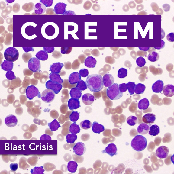
    Episode 209: Blast Crisis May 01, 2025
    Show notes

    We dive into the recognition and management of blast crisis.

    Hosts:
    Sadakat Chowdhury, MD
    Brian Gilberti, MD

    https://media.blubrry.com/coreem/content.blubrry.com/coreem/Blast_Crisis.mp3 Download 2 Comments Tags: Hematology, Oncology

    Show Notes

    Topic Overview

    • Blast crisis is an oncologic emergency, most commonly seen in chronic myeloid leukemia (CML).
    • Defined by:
      • >20% blasts in peripheral blood or bone marrow.
      • May include extramedullary blast proliferation.
    • Without treatment, median survival is only 3–6 months.

    Pathophysiology & Associated Conditions

    • Usually occurs in CML, but also in:
      • Myeloproliferative neoplasms (MPNs)
      • Myelodysplastic syndromes (MDS)
    • Transition from chronic to blast phase often reflects disease progression or treatment resistance.

    Risk Factors

    • 10% of CML patients progress to blast crisis.
    • Risk increased in:
      • Patients refractory to tyrosine kinase inhibitors (e.g., imatinib).
      • Those with Philadelphia chromosome abnormalities.
      • WBC >100,000, which increases risk for leukostasis.

    Clinical Presentation

    • Symptoms often stem from pancytopenia and leukostasis:
      • Anemia: fatigue, malaise.
      • Functional neutropenia: high WBC count, but increased infection/sepsis risk.
      • Thrombocytopenia: bleeding, bruising.
    • Leukostasis/hyperviscosity effects by system:
      • Neurologic: confusion, visual changes, stroke-like symptoms.
      • Cardiopulmonary: ARDS, myocardial injury.
      • Others: priapism, limb ischemia, bowel infarction.
    • Rapid deterioration is common — early recognition is critical.

    Diagnostic Workup

    • CBC with differential: assess blast % and cytopenias.
    • Peripheral smear and manual diff: confirm immature blasts.
    • CMP: screen for tumor lysis syndrome:
      • Elevated potassium, phosphate, uric acid.
      • Low calcium.
    • LDH & uric acid: markers of high cell turnover.
    • Coagulation studies (PT, PTT): assess for DIC.
    • Definitive tests (done inpatient): bone marrow biopsy, flow cytometry.

    Emergency Department Management

    • Resuscitation & ABCs: oxygen, IV fluids, vitals monitoring.
    • Avoid aggressive transfusions:
      • Risk of hyperviscosity with PRBCs and platelets.
    • Initiate broad-spectrum antibiotics early:
      • High suspicion for sepsis in functionally neutropenic patients.
    • Consider antifungals for prolonged febrile neutropenia.
    • Cytoreduction strategies:
      • Hydroxyurea to lower WBCs quickly.
      • Tyrosine kinase inhibitors (TKIs).
      • High-dose chemotherapy.
    • Early consultation with hematology/oncology is essential.
    • Mutation testing may guide targeted therapy.

    Prognosis

    • Without treatment: median survival ~3 months.
    • With treatment:
      • Potential survival >1 year.
      • Best outcomes in patients who enter a second chronic phase and undergo allogeneic stem cell transplant.

    Ethical & Logistical Considerations

    • Treatment may involve aggressive interventions with serious side effects.
    • Important to assess:
      • Patient goals of care.
      • Capacity for informed consent.
    • Resource limitations:
      • Not all hospitals have oncology services.
      • Patients may require transfer over long distances.
    • Emphasize early, transparent discussions with patients and families.

    Top 3 Take-Home Points

    • Recognize early: Look for cytopenias, leukostasis, and rapid clinical decline.
    • Resuscitate appropriately: Start antibiotics; be cautious with transfusions.
    • Call for help: Early hematology/oncology involvement is essential for definitive care.

    Read More

    Episode 208: Geriatric Emergency Medicine Apr 15, 2025
    Show notes

    We explore the expanding field of Geriatric Emergency Medicine.

    Hosts:
    Ula Hwang, MD
    Brian Gilberti, MD

    https://media.blubrry.com/coreem/content.blubrry.com/coreem/Geriatric_Emergency_Medicine.mp3 Download One Comment Tags: Geriatric

    Show Notes

    Key Topics Discussed

    • Importance and impact of geriatric emergency departments.
    • Optimizing care strategies for geriatric patients in ED settings.
    • Practical approaches for non-geriatric-specific EDs.

    Challenges in Geriatric Emergency Care

    • Geriatric patients often present with:
      • Multiple chronic conditions
      • Polypharmacy
      • Functional decline (mobility issues, cognitive impairments, social isolation)

    Adapting Clinical Approach

    • Core objective remains acute issue diagnosis and treatment.
    • Additional considerations for geriatric patients:
      • Review and caution with medications to prevent adverse reactions.
      • Address functional limitations and cognitive impairments.
      • Emphasize safe discharge and care transitions to prevent unnecessary hospitalization.

    Identifying High-Risk Geriatric Patients

    • Screening tools:
      • Identification of Seniors at Risk (ISAR)
      • Frailty screens
    • Alignment with the “Age-Friendly Health Systems” initiative focusing on:
      • Mentation
      • Mobility
      • Medications
      • Patient preferences (what matters most)
      • Mistreatment (elder abuse awareness)

    Minimizing Hospital-Related Harms

    • Involvement of multidisciplinary teams:
      • Social workers and care managers for care transitions
      • Geriatric-certified pharmacists for medication review
    • Coordination with outpatient services post-discharge

    Implementing Geriatric Care in All EDs

    • Basic geriatric care achievable even in resource-limited or rural EDs.
    • Level 3 Geriatric ED Accreditation can be achieved through:
      • Improved care transitions
      • Staff education enhancements
      • Age-friendly environments (comfort, nutrition, hydration)

    Future of Geriatric Emergency Medicine

    • Vision: Universal integration of geriatric-focused care.
    • Goals:
      • Enhanced patient experience
      • Improved care transitions
      • Alignment of treatments with patient goals
      • Broader enhancement of emergency care quality for all patient populations

    Read More

    Previous 1 2 3 4 24 Next

    Related Podcasts

    The Joe Rogan Experience

    1

    The Joe Rogan Experience Comedy
    More Perfect

    2

    More Perfect Government
    Casefile True Crime

    3

    Casefile True Crime Games & Hobbies
    Reply All

    4

    Reply All Games & Hobbies
    You Must Remember This

    5

    You Must Remember This Games & Hobbies
    Real Time with Bill Maher

    6

    Real Time with Bill Maher Games & Hobbies
    footer-logo

    Contact Us

    Toll Free: 844-670-7747

    Links

    • Home
    • Top Charts
    • Networks
    • Apps
    • Independents Podcasts
    • Podcast Advertising
    • Podcast News
    • Contact Us
    • About Us
    • Analytics & Insights

    Stay Connected

      Privacy, Terms of Use & Our Code of Ethics Protecting Content Creators Copyrights