An evidence-based emergency medicine podcast. The Washington University Emergency Medicine Journal Club podcast provides an excellent way to keep up with the medical literature, improve patient care, and increase understanding of evidence-based medicine. Each month we ask a clinical or operational question pertinent to the practice of emergency medicine. We then discuss the four articles most relevant to that question in an attempt to determine the most evidence-based approach to patient care.
It is 0200 and you are working in the trauma critical care (TCC) area of your beloved emergency department. During the chaotic shift, three patients require endotracheal intubation. The first patient is a 55 year old man with a history of lymphoma in remission, and he is status post splenectomy. He arrived hypoxic with a SpO2 of 85% and tachypneic with a respiratory rate of 30. It is obvious what is needed and he is immediately intubated successfully without complication in room 5 of the TCC. His post-intubation chest radiograph shows bilateral alveolar opacities congruent with multi-focal pneumonia, and acute respiratory distress syndrome (ARDS).
You walk out of room 5 and see an obtunded 22 year female vomiting in room 6. She is intoxicated and smells of alcohol, and a quick biopsy of her belongings shows several prescription bottles. You think this is likely a polysubstance overdose, and because of her mental status and lack of gag, you intubate for airway protection. Vomitus is around her vocal cords during direct laryngoscopy and she has significant secretions noted by the respiratory therapist. Her post-intubation chest radiograph shows a right lower lobe infiltrate, most congruent with aspiration.
As you exit room 5, you are called to room 4 because of the arrival of a 45 year old man who was trying to light his cigarette while bent over a brewing batch of methamphetamines. Shockingly, a small eruption occurred. Upon arrival, he is alert (also intoxicated) and you note that he has minimal burns, all located to his face, with singed hairs and soot in his mouth. You perform a quick fiberoptic examination of his upper airway, and note edematous and erythematous tissue in his posterior oropharynx, extending distally to his vocal cords. Given this inhalation injury and potential for upper airway obstruction, you intubate him, which is uneventful. His post-intubation chest radiograph is remarkable only for some interstitial edema.
You are happy that you have successfully performed perhaps the most defining trait of a well-trained emergency physician- securing an emergent airway. Your respiratory therapist is asking for ventilator orders, and she has set each patient at what appears to be pretty standard settings, with a tidal volume of 500mL for each patient. Noting that the median ICU wait time at your institution is around 6 hours, you wonder if this catch-all mechanical ventilation prescription is the most appropriate strategy. You are now considering the evidence regarding tidal volume for patients with ARDS, but especially for the two patients at risk for ARDS.
PICO Question:
Population: Mechanically ventilated adult patients at risk for ARDS, but without the syndrome.
Intervention: Low tidal volume (≈6mL/kg PBW).
Comparison: Conventional tidal volume.
Outcome: Incidence of ARDS early after ICU admission from the ED (i.e. within ≈5 days).
Search Strategy:
MEDLINE, EMBASE, CINHAL, and the Cochrane Library were searched using:
Acute Lung Injury
"Acute Lung Injury"[Mesh] OR "Acute Lung Injury" OR “Acute Lung Injuries” OR "Ventilator-Induced Lung Injury"[Mesh] OR "Ventilator-Induced Lung Injury” OR “Ventilator-Induced Lung Injuries” OR “Ventilator Associated Pneumonia” OR “ventilation induced lung injury” OR “VILI”
AND
Prevention
Prevent* OR prevention OR prophylax* OR prophylac* OR chemoprevent* OR thwart* OR "ward off" OR "ward-off" OR pre-emptive* OR preemptive* OR chemoprophyla*
AND
Outcome
(outcome* OR ((treatment* OR protocol*) AND (respond* OR response*)) OR failure* OR mortality OR fatal* OR death OR dead OR deaths OR "passed away" OR demise* OR Recurren* OR progression OR progressed OR relaps* OR growth OR grew OR growing OR regression OR survival OR cure OR cures OR "quality of life" OR qol OR morbidit* OR adverse OR "side effect" OR "side effects" OR event OR events OR nausea OR nauseous OR vomit* OR emesis OR comfort* OR pain OR painful OR painfree OR pain-free OR stress OR analges* OR "Outcome Assessment Health Care "[Mesh] OR "Mortality"[Mesh] OR "mortality "[Subheading] OR "Survival"[Mesh] OR "Survival Analysis"[Mesh] OR "Quality of Life"[Mesh] OR "Pain Measurement"[Mesh] OR "Health"[Mesh] OR "Health Status Indicators"[Mesh] OR "Health Status"[Mesh] )
AND
Adults
"Young Adult"[Mesh] OR “Young adults” OR “Young Adult” OR "Adult"[Mesh] OR “adults” OR “adult” OR "Middle Aged"[Mesh] OR “Middle age” OR "Aged"[Mesh] OR “Aged” OR Elder* OR "Aged, 80 and over"[Mesh] OR “Oldest Old” OR Nonagenarian* OR Octogenarian* OR Centenarian* OR "Frail Elderly"[Mesh] OR “Frail Older Adults” OR “Frail Older Adult”
AND
PubMed NOT Animal Studies
NOT (("Animals"[Mesh]) NOT ("Animals"[Mesh] AND "Humans"[Mesh]))
This yielded a total of 1,704 potentially relevant articles, of which 1,652 were excluded based on title on abstract. This left 52 full text articles, which were further reviewed to assess appropriateness and relevance to the journal club topic.
Bottom Line:
With an incidence of close to 200,000 patients annually (Rubenfeld 2005), a mortality rate of about 40% (Bersten 2002, Rubenfeld 2007), and prolonged physical and mental sequelae in survivors, it is clear that ARDS is one of the most important problems in critical care. A treatment still does not exist for the underlying pathophysiology of the syndrome, and low tidal volume ventilation remains the only consistent mortality benefit across syndrome severity. Given these facts, prevention of ARDS has become increasingly important, and the focus of increasing study and funding priorities.
A major question, and topic of controversy, is “Should lower tidal volume be used prophylactically to prevent ARDS in at-risk patients?” Tidal volume is a major risk factor for the development of ventilator-associated lung injury (VALI). This has been established in animals models of lung injury and human data in patients with ARDS.
ARDS is relatively common in the ED. Observational data and a small randomized controlled trial (Determan 2010) show that lower tidal volume may reduce progression to ARDS. This was further studied by two recently published systematic reviews (Serpa Neto 2012, Fuller 2013), which both concluded that the majority of data does point to a therapeutic benefit to low tidal volume ventilation to prevent ARDS.
Another major question is “Can the ED play a role in the treatment and prevention of ARDS?” Observational data shows that a significant minority of patients have ARDS in the ED (Fuller 2013). Clinical data also shows that progression to ARDS occurs early after ICU admission, with a median onset of 30 hours (Shari 2011). This temporal relationship suggests a potential causal link to treatment provided (or not provided) during the ED and early ICU course. Despite this, not a single trial of mechanical ventilation has ever been conducted in the ED. Further highlighting the importance of ARDS prevention, patients progressing to ARDS experience increased morbidity and mortality.
With respect to mechanical ventilation in the ED, there is limited data. What does exist tells us that tidal volumes commonly delivered are highly variable, and potentially injurious ventilation practices exist. This provides rationale for quality improvement, knowledge translation, and randomized trials in these ED patients.
The HINTS Exam in Vertigo
Feb 02, 2014
Show notes
Journal Club Podcast #10: January 2014 Dr. David Newman-Toker, the authority on the HINTS exam, joins me to talk about oculomotor testing in acute vertigo... Download the Podcast Central Vertigo Video Links:A HINTS exam consistent with vertigo of central should have at least one of the following: a normal head impulse test (without a corrective saccade), nystagmus that changes direction on eccentric gaze, or a positive test of skew deviation (vertical ocular misalignment). Normal head impulse test Direction-changing nystagmus Positive test of skew - Courtesy of Dr. Peter Johns Peripheral Vertigo Video LinksA HINTS exam consistent with peripheral vertigo should have all of the following: an abnormal head impulse test (with a corrective saccade), nystagmus that does not change direction on eccentric gaze, and a negative test of skew deviation. Abnormal head impulse test - Courtesy of Dr. Peter Johns Unidirectional nystagmus - Courtesy of Dr. Kevin Kerber Negative test of skew Click Tab to Expand Articles: Article 1: Newman-Toker DE, Kerber KA, Hsieh YH, Pula JH, Omron R, Saber Tehrani AS, Mantokoudis G, Hanley DF, Zee DS, Kattah JC. HINTS Outperforms ABCD2 to Screen for Stroke in Acute Continuous Vertigo and Dizziness. Acad Emerg Med. 2013 Oct;20(10):986-996. Answer Key. Article 2: Kattah JC, Talkad AV, Wang DZ, Hsieh YH, Newman-Toker DE. HINTS to diagnose stroke in the acute vestibular syndrome: three-step bedside oculomotor examination more sensitive than early MRI diffusion-weighted imaging. Stroke. 2009 Nov;40(11):3504-10. Answer Key. Article 3: Newman-Toker DE, Kattah JC, Alvernia JE, Wang DZ. Normal head impulse test differentiates acute cerebellar strokes from vestibular neuritis. Neurology. 2008 Jun 10;70(24 Pt 2):2378-85. Answer Key. Article 4: Newman-Toker DE, Saber Tehrani AS, Mantokoudis G, Pula JH, Guede CI, Kerber KA, Blitz A, Ying SH, Hsieh YH, Rothman RE, Hanley DF, Zee DS, Kattah JC. Quantitative video-oculography to help diagnose stroke in acute vertigo and dizziness: toward an ECG for the eyes. Stroke. 2013 Apr;44(4):1158-61. Answer Key. Vignette: While moonlighting in a small, community hospital one evening, you are presented with a 58 year-old gentleman complaining of vertigo. He was at home eating dinner 5 hours prior to arrival when he felt the room begin to suddenly, and violently, spin around him. He notes “I haven’t felt like this since college!” He reports becoming nauseated and vomiting several times, then getting up and “staggering” to his bed where he laid down and tried to “wait it out.” After several hours of constant vertigo, he attempted to get up to go to the bathroom and fell to the floor. He managed to grab his cellphone and called 911. He reports the vertigo has been constant, is worse with any change in head position, and is associated with nausea and imbalance. He denies recent URI symptoms, hearing changes, focal weakness or numbness, or speech changes. His past medical history includes hypertension and diabetes, controlled with amlodipine, metformin, and glyburide. On exam, he has horizontal beating nystagmus. Cerebellar exam - including finger to nose, heel to shin, and rapid alternating movements - is otherwise normal. He has an abnormal Romberg’s and is unable to stand or ambulate unassisted. The remainder of his neurologic exam is normal. Head CT, ECG, and labs are all normal. Your differential includes two main concerns: either the patient has vestibular neuritis and should be treated symptomatically and discharged, or he has suffered a cerebellar stroke and requires transfer to a hospital with neurology consultation and MRI available. When the patient does not improve after receiving oral meclezine and IV diazepam, you bite the bullet and transfer him to Barnes-Jewish for further evaluation by the stroke team. On your way home the next morning, you begin wondering if there are any aspects of the physical exam that can differentiate between peripheral and central causes of vertigo. A quick search of the literature identifies something referred to as the “HINTS” exam, which involves oculomotor testing. Specifically, this test includes evaluation of horizontal Head Impulse testing, the direction of Nystagmus, and Test of ocular Skew deviation. You begin delving deeper to determine if this is something you should be using in your practice… PICO Question: Population: Adults with new-onset, acute vertigo with otherwise non-focal neurologic exam Intervention: Bedside oculomotor testing (HINTS exam) Comparison: MRI, discharge diagnosis, follow-up diagnosis Outcome: Diagnostic accuracy, morbidity or mortality related to misdiagnosis Search Strategy: An advanced PubMed search was conducted using the terms "(HINTS OR oculomotor OR vestibuloocular) AND (vertigo or dizziness)," limited to humans and the English language, resulting in 142 citations (http://tinyurl.com/mktaevm). Original studies that reported sufficient data to construct 2X2 contingency tables were chosen for analysis. The bibliographies of relevant articles were searched for additional references. Three articles that specifically addressed the diagnostic accuracy of the HINTS exam were identified. An additional article was selected that assessed the 3 components of HINTS as well as vertical smooth pursuit, but allowed for calculation of the accuracy of the HINTS exam alone. Bottom Line: Dizziness remains a common chief complaint in US emergency departments, leading to approximately 4 million visits every year (Saber Tehrani 2013). The emergency physician’s first duty in such cases is to distinguish benign peripheral causes of vertigo from more serious, potentially life-threatening, central causes. Making such a determination can be difficult: focal neurologic signs are absent in as many as 20% of cases of posterior circulation stroke (Tarnutzer 2011); computed tomography (CT) is frequently normal early in the course of posterior circulation stroke (Edlow 2008); and magnetic resonance imaging (MRI), often considered the reference standard for stroke, is associated with a significant number of false negatives when the posterior circulation is involved (Oppenheim 2000, Morita 2011). Emergency physicians have identified the importance of a clinical decision rule to help differentiate central from peripheral etiologies of vertigo (Eagles 2008). The HINTS exam has been proposed as a means of making such a differentiation. This test involves 3 components: 1) Horizontal head impulse testing involves rapid head rotation by the examiner with the subject’s vision fixed on a nearby object (often the examiner’s nose). In cases of peripheral vertigo, a corrective saccade should be observed, and is considered a positive test. There is typically no corrective saccade in cases of central vertigo. 2) Evaluation of nystagmus will typically yield a fast phase which is unidirectional in peripheral vertigo, and beats away from the affected side. In central vertigo, the direction of the fast phase may change on eccentric gaze. 3) Alternate eye cover testing in patients with peripheral vertigo should result in no skew deviation or ocular tilt. Ocular misalignment and skew deviation (with or without ocular tilt) is frequently seen in patients with posterior fossa abnormalities (i.e. brainstem strokes). Download the HINTS video demonstration While its individual components do not reliably differentiate central from peripheral causes of vertigo, an exam consisting of all three elements has been proposed to do so. In theory, if any of the components indicates a central pathology, then the exam is considered positive for a central etiology. All three components must be consistent with a peripheral etiology for the exam to be considered negative. The current literature supporting the HINTS exam consists of four articles, three of which included patients from a single, ongoing prospective cross-sectional diagnostic study of patients with acute vestibular syndrome (AVS). The first article (Kattah 2009) included 101 patients with acute vertigo, of whom 76 were diagnosed with a central lesion. The diagnostic test characteristics of the HINTS exam for central vertigo were as follows: the sensitivity was 100% (95% CI 95.2-100.0), specificity was 96% (95% CI 79.6-99.3), likelihood ratio positive (LR+) was 25 (95% CI 3.66 to 170.59), and LR negative (LR-) was 0.00 (95% CI 0.00 to 0.11). Interestingly, the HINTS exam outperformed the initial MRI with diffusion-weighted imaging, which had a sensitivity for stroke of only 88%. Similar diagnostic properties were identified in the 2nd paper (Newman-Toker 2013) which compared the accuracy of the HINTS exam to the ABCD2 score in 190 patients from the cross-sectional cohort. The ABCD2 score is a clinical prediction rule to predict short-term stroke risk following a transient ischemic attack. While this comparison seems contrived and unfair, the HINTS test performed quite well, with a sensitivity of 96.8% (95% CI 92.4-99), a specificity of 98.5% (95% CI 92.8-99.9), a LR+ of 63.9 (95% CI 9.13-446.85), and LR- of 0.03 (95% CI 0.01-0.09). The 3rd study from this database (Newman-Toker 2013) used a small sample of 12 patients to evaluate the HINTS exam aided by a video-oculography device, which was used to record head and eye velocity measurements during head impulse testing. Physician interpretation of these reading was compared to an algorithmic interpretation, with 100% agreement. The aided HINTS exam demonstrated a high-degree of accuracy in the diagnosis of central vertigo, with a sensitivity and specificity of 100% (95% CI 54.1-100.0%), LR+ of ∞, and LR- of 0. A 4th article was identified in which oculomotor testing was performed by neurologists, following completion of 4 hours of training specific to exam techniques and interpretation. Twenty-four patients admitted to the stroke unit were included in the study, of whom 10 were diagnosed with central vertigo. The sensitivity of the HINTS exam for stroke was 100% (95% CI 69.0 to 100.0), specificity was 85.7% (95% CI 57.2-97.8), LR+ was 7.0 (95% CI 1.9 to 25.3), and LR- was 0. Several concerns were raised with regards to the current evidence. First, in these studies the HINTS exam was performed by specialists: neuro-ophthalmologists in two studies, neuro-otologists in one study, and neurologists with four hours of exam-specific training in the 4th study. This calls into question the external validity of the study, as the accuracy and reliability of HINTS testing in the hands of emergency physicians has not been evaluated. However, with some degree of training it is reasonable to expect emergency physicians to be able to perform the HINTS exam as proficiently as our specialist colleagues. When ED ultrasound was first being introduced, one of the primary concerns was that "only trained radiologists" could perform ultrasound and hence this was out of the jurisdiction of the ED. Over the years, we have not only proven capable of using ultrasound effectively in emergency medicine, but ultrasound is now a requirement of emergency medicine residency training. The question will be whether the amount of training necessary to become proficient with the HINTS exam will be worth the effort. A second concern raised was that the patient populations in these studies were of moderate to high risk of central vertigo, with prevalence ranging from 42 to 75%. While the prevalence was high in all of these studies, these were still fairly heterogeneous groups of patients with variable risk (age ranges of 18-92, 26-92, 42-83, and 30-73) and hence did include some patients we would likely consider low risk. Some of the patients with stroke as the cause of symptoms were young (15 patients < 50 years of age in the study by Kattah et al). I would argue that some of these patients with central lesions would be treated as peripheral vertigo and discharged without advanced imaging at most institutions, and an abnormal bedside test would potentially lead to admission and further testing, and reduce the rate of missed stroke. In patients with low probability of disease, an abnormal HINTS exam may increase the pre-test probability of disease above the test threshold for MRI or admission. This is especially true at institutions without MRI or neurologic consultation available, where transfer to another hospital for admission and further work-up would be required to assess for a central etiology. In some moderate risk patients, a negative HINTS exam may reduce the probability of central etiology below the test threshold, and obviate the need for further work-up. For example, using the upper limits of the 95% CI for the negative LR from the largest study (Newman-Toker 2013) of 0.09, a patient with a pre-test probability of 25% for a central etiology who has a negative HINTS would have a post-test probability of 2.9%, and hence the decision may be made to not proceed with further work up. Further testing of the HINTS exam will need identify a more concrete role for this test. The accuracy and reliability of the test in the hands of trained emergency physicians will need to be assessed, as will the impact of the test on both cost and patient care. If use of the test does not lead to either reduction in unnecessary imaging, reduction in the rates of missed posterior circulation infarction, or both, then it will not be worth the effort to train physicians in its performance. The role of the video-oculography device will also need to be further assessed in larger studies with more precise estimates of diagnostic accuracy to justify its cost.
Pediatric Volume Resuscitation in the Developing World
Dec 04, 2013
Show notes
Journal Club Podcast #8: November 2013
Chris Carpenter sits down with Indi "we named the dog Indiana" Trehan and SueLin Hilbert to talk about pediatric volume resuscitation in the developing world...
The infamous SueLin Hilbert has brought you to Ghana to help staff the emergency department and train local Ghanaian physicians in the art and science of emergency medicine. Aside from the local beer, you are loving the experience and you are making a big positive impact on clinical care in this very underserved population. One night, you are covering the ED alone when a 4-year-old girl arrives in septic shock. She is febrile, hypotensive, tachycardic, barely responsive to painful stimulation, and has delayed capillary refill. You are not sure what the underlying diagnosis is and cannot find a specific diagnosis due to the fact that it is the middle of the night and the malaria lab techs are gone for the night and you are out of rapid malaria diagnostic tests. But, it is the rainy season and you are seeing tons of malaria, so you assume the child has malaria sepsis most likely or less likely Gram-negative bacterial sepsis.
Nevertheless, it is clear to you she is in septic shock. You manage to place an EJ line after the nurses fail to obtain peripheral access and you aggressively resuscitate her with normal saline at a dose of 60 mL/kg over half an hour, broad-spectrum antibiotics, and supplementary oxygen. You consider albumin and blood transfusions, but by the time you get back to this patient (since you have an ED full of other sick patients), you learn that her respiratory status worsened, she became more pale and cold, and ultimately died. Her mother is shrieking and crying. You are at the edge of tears since this is certainly not the first child that has died on your watch today.
In the morning after your shift, you go home, have a stiff drink, and ponder what you may or may not have done wrong. After discussing it with SueLin in the morning, she makes you think about whether the choice and volume of fluids you administered were ideal. You get on Pubmed to consider the role of fluid resuscitation in septic shock in the developing world and come across the following articles:
PICO Question:
Population: Children with septic shock due to severe infection in the developing world.
Intervention: Fluid resuscitation with alternate fluids or rates.
Comparison: Standard resuscitation with 20 mL/kg normal saline.
Outcome: Survival and time to recovery from shock.
Search Strategy:
PubMed was searched on October 11, 2013, using the strategy: (sepsis or malaria) AND (fluid resuscitation). This gave 1182 results. The “English” language filter was applied. This reduced the total to 1064 results. The “child” age filter was applied. This reduced the total to 169 results. The “10 years” publication date filter was applied. This reduced the total to 85 results (http://goo.gl/cdn1r1). The abstracts of these 85 articles were reviewed to assess for relevant studies conducted in the developing world.
Bottom Line:
Circulatory shock is a significant problem in pediatric emergency medicine and critical care. The leading culprit is hypovolemia, usually due to life-threatening infection. Consequently, current guidelines for the acute management of severe sepsis in pediatric (and adult) patients emphasize early, rapid, and substantial infusion of intravenous fluids. The optimal fluid choice, volume of fluid, and route of administration has been the topic of debate for 175 years. Indeed, this topic was the focus of at least two recent systematic reviews. (Akech 2010, Ford 2012) Nonetheless, this remains a contentious issue as evidenced by numerous letters and commentaries. (Ford 2011, Ribeiro 2011, Berend 2011, Joyner 2011, Kissoon 2011, Scott 2011, Myburgh 2011, Hilton 2012)
The highest-quality (least biased) evidence to date is the Fluid Expansion as Supportive Therapy in Critically Ill African Children (FEAST) trial, although thus far no guideline revisions reflect the findings of this study. Although the FEAST trial represents landmark research, global medicine experts at our Journal Club identified several potentially serious flaws with it. First, the trial closed prematurely (with pros and cons), although the decision appears justified on ethical grounds. The authors provided much more detail about this decision in a subsequent manuscript. Second, the investigators failed to provide or analyze the cause of death. One Journal Club attendee noted that a webinar conducted by several of the FEAST site investigators analyzing these findings identified “cardiac collapse” as the cause of death in most of these children. Identifying the cause of death will be important to guide subsequent management trials, as well as to fully understand the implications of this FEAST trial. Third, the authors failed to use the World Health Organization criteria for shock since the role of physical exam to stratify severity of illness in normotensive children is unproven. Whereas the WHO requires the presence of delayed capillary refill, weak pulse, and tachycardia to establish the diagnosis of “shock”, these investigators only required one of the three. Were the children in this FEAST trial septic shock patients? One editorial suggests that FEAST was “probably treating children with serious febrile illnesses due to the most common medical problems, namely pneumonia and malaria, but not hypovolaemic shock.”
What did the FEAST trial conclude? Routine IVF bolus therapy in clinically undifferentiated severely ill non-hypotensive febrile children with diminished perfusion increases 24-hour mortality whether normal saline or albumin is used. Increased mortality occurs regardless of malaria status, coma, severe anemia, base deficit, or lactate level. Hypothetical mechanisms include non-blood product fluid resuscitation in severe anemia, rapid reversal of compensatory vasoconstrictor response, reperfusion injury, subclinical pulmonary compliance effects, sepsis-relatedmyocardial function, or intracranial pressure. Before extrapolating the FEAST findings to the developed world, future research would need to explore similar fluid resuscitation strategies in the context of readily available ICU and mechanical ventilation.
The rest of the evidence that we evaluated was of lesser quality and more focused on specific disease processes (malaria) or fluid types (Dextran vs. hydroxyl ethyl starch vs. normal saline or lactated ringers). The PGY-1 manuscript demonstrated that in pediatric patients with severe malaria and moderate to severe acidosis in the developing world, IV albumin at 20-40 cc/kg plus standard antimicrobial and supportive therapy is superior to IV normal saline with an adjusted Number Needed to Treat (NNT) of 3 to prevent one death based upon a baseline mortality rate of 11%. Albumin is more effective in malaria patients presenting with coma.
The PGY-2 manuscript demonstrated that in children with shock in India, more aggressive fluid and dopamine resuscitation within the first 20 minutes of ED arrival does not decrease mortality or increase intubation rates. If clinical equipoise remains despite these findings, future researchers should evaluate settings with >1 ED clinician and more ready access to ventilators to more accurately assess the internal and external validity of this intervention.
The PGY-3 manuscript demonstrated that in severe (impaired consciousness or respiratory distress) pediatric malaria in the developing world, hydroxyl ethyl starch (HES) and Dextran are both safe for acute volume expansion therapy and no adverse outcomes observed among 80 patients. However, impressive trends were observed favoring HES (compared with Dextran) to reduce mortality and resolve acidosis at eight hours with NNT=5.
The consensus of our Pediatric EM and Critical Care Medicine experts was that the lack of access to ventilator support and Intensive Care Unit management in the FEAST trial limit prohibit extrapolation of these findings to the developed world at places like St. Louis Children’s Hospital. As far as the experts were aware, no ongoing or future studies were planned to further evaluate the efficacy of aggressive fluid resuscitation in pediatric septic shock patients and neither guidelines nor local protocols would change based upon the FEAST trial. Nonetheless, an expanding volume of research in the developed world using contemporary ICU management indicates that excessive fluid resuscitation contributes to preventable morbidity including sepsis, ARDS, acute kidney injury, and major surgery. Several ongoing trials will assess the effectiveness of early goal directed therapy (which includes aggressive fluid resuscitation) in septic shock management. However, Washington University School of Medicine global medicine experts Dr. Indi Trehan and Dr. SueLin Hilbert noted that the FEAST trial radically altered the approach to septic shock resuscitation in the developing world in that undifferentiated rapid fluid resuscitation is no longer the standard of care.
One unintended consequence of this Pediatric EM-Pediatric Critical Care-Emergency Medicine Journal Club event was recognition of a potential niche in diagnostic research. Specifically, the reproducibility of bedside features of shock (and of history and physical exam in most of Pediatrics, as well as systematic reviews of diagnostic accuracy) is under-researched, largely unknown and poorly reported. How accurate and reliable are pediatric clinicians’ physical exam skills for detecting “severe sepsis”, organomegaly, etc.? This represents a tremendous opportunity (i.e. unfilled academic niche) for diagnostic researchers to contribute to the JAMA Rational Clinical Exam and Academic Emergency Medicine Evidence Based Diagnostics series.
Cardiac Cath Following Cardiac Arrest in Patients Without STEMI
Nov 11, 2013
Show notes
Journal Club Podcast #7: October 2013
This month, the outspoken Chandra Aubin and hold hands and talk about which patients should undergo urgent cardiac cath following cardiac arrest...
While on your EMS rotation, you ride along on a call for a 52-year old gentleman with cardiac arrest. On arrival to the patient's home, bystander CPR is underway. You learn that 10 minutes prior to your arrival, the patient was running on his treadmill when he suddenly clutched his left shoulder. He turned off the treadmill, told his wife and daughter he didn't feel well, and collapsed to the floor. His 17-year old daughter (a lifeguard) attempted to feel for a pulse while his wife called 911. The daughter was unable to palpate a pulse and began CPR.
The paramedics continue CPR while you attach the cardiac monitor. Seeing that the rhythm is ventricular fibrillation, you place the pads and defibrillate the patient with success on the first attempt. He now has a pulse, and his BP is 85/40 with a pulse of 115. After placing an IV and intubating the patient, you then transport him to the Barnes-Jewish ED. His ECG on arrival reveals sinus tachycardia with normal intervals and deep T-wave inversions in leads V1-3, with no ST elevation. His BP is now up to 110/60, and his pulse remains 115. The ED team begins placing an ICY line to induce mild therapeutic hypothermia, sends labs, and begins the process of admission to the ICU.
You feel fairly certain, based on the history provided by family, that the underlying cause of this patient's cardiac arrest was an acute myocardial infarction. Despite the lack of ST-elevation the ECG, you wonder if the patient would benefit from early cardiac catheterization to assess for significant acute coronary artery occlusion. You head to the offices on the 8th floor, find a computer, and begin a search to see what the literature says...
PICO Question:
Population: Adults patients with out-of-hospital cardiac arrest who achieve return of spontaneous circulation without evidence of STEMI on the initial ECG (with ventricular arrhythmia vs. any rhythm).
Intervention: Early cardiac catheterization.
Comparison: Standard care, delayed cardiac catheterization, no cardiac catheterization.
Outcome: Findings of acute coronary artery occlusion, survival to hospital discharge, good neurologic recovery, quality of life.
Search Strategy:
PubMed was searched using the strategy "(cardiac arrest) AND (early OR immediate) AND ((cardiac catheterization) OR (coronary angiography) OR (coronary intervention))" (http://tinyurl.com/lke4so9), resulting in 602 articles, from which the following 4 articles were chosen.
Bottom Line:
A large meta-analysis of data on outcomes from out-of-hospital cardiac arrest (OHCA) revealed that less than one-third of patients with return of spontaneous circulation (ROSC) survive to hospital discharge (Sasson 2010). While this study pre-dates the use of interventions shown to improve survival with good neurologic outcome, such as therapeutic hypothermia (Holzer 2005, Kim 2012), the data does suggest the need for improvements in the care of post-arrest patients. The 2010 American Heart Associations Guidelines for Post-Cardiac Arrest Care describe therapeutic hypothermia and “treatment of the underlying cause of cardiac arrest” as the primary initiatives to improve survival and neurologic outcome. Acute myocardial infarction has been demonstrated to be the likely cause of arrest in anywhere from one-third (Spaulding 2007, Anyfantakis 2009) to 61% (Chelly 2012) of patients admitted to the hospital. In patients with ST-elevation myocardial infarction (STEMI) on ECG following ROSC, the International Liaison Committee on Resuscitation (ILCOR) clearly recommends that reperfusion therapy be attempted. In patients without STEMI, ILCOR is less clear, though, and recommends only that one “consider immediate coronary angiography in all post-cardiac arrest patients in whom ACS is suspected.”
One systematic review looked at studies on coronary angiography in OHCA (Larsen 2012). A meta-analysis of 10 studies in which coronary angiography was performed in select patients revealed an unadjusted odds ratio for survival of 2.78 (95% CI 1.89-4.10). While this finding suggests improved outcomes with coronary angiography, selection bias likely made a large contribution given the selective, observational nature of these studies. The authors also identified 5 studies that assessed the use of coronary angiography systematically in all survivors of OHCA without an obvious non-cardiac cause, and found that the presence of significant coronary artery disease was high, ranging from 59-71%, despite only 31-63% of included patients presenting with STEMI or presumed new LBBB. This suggests that a significant subset of patients without STEMI had significant coronary occlusion, and could potentially benefit from PCI. While this systematic review suggests that there is a subset of patients in whom coronary occlusion is the underlying cause for the arrest, but in whom STEMI is absent on the post-resuscitation ECG, it is unclear which patients will benefit from attempts at coronary reperfusion.
In the Parisian healthcare system, all patients successfully resuscitated after OHCA are transported immediately for coronary angiography (Laurent 2002, Dumas 2010, Chelly 2012). Dumas identified 435 patients with ROSC following OHCA without an obvious non-cardiac etiology. Among patients with STEMI, 96% had at least one significant coronary stenosis on angiography; in patients without STEMI, 58% had at least one significant coronary stenosis. While this latter number would indicate that cardiac catheterization was necessary in the majority of such patients, it is important to note that only 26% of these patients underwent successful percutaneous coronary intervention (PCI). The authors primary outcome was survival in patients undergoing successful PCI compared to those with no or failed PCI; they found survival rates of 51% vs. 31% respectively (p < 0.001) for a relative risk of 1.62. The authors’ conclusion, that “immediate PCI seems to offer survival benefit” (p. 206) fails to address one important concern: the difficulty in predicting successful PCI in non-STEMI patients prior to coronary angiography. A more accurate conclusion may be that if you are going to have a cardiac arrest, make sure you have a lesion amenable to PCI.
In an attempt to reduce the rates of unnecessary cardiac catheterization, another Parisian study attempted to identify ECG findings predictive of angiographically defined acute myocardial infarction (AMI) in OHCA patients (Sideris 2011). They found that the combination of ST-elevation, ST-depression, left bundle branch block (LBBB), or nonspecific QRS widening had a sensitivity of 100%, a specificity of 47%, a positive predictive value of 52%, and a negative predictive value of 100%. Of 46 patients without ST-elevation who were positive by ECG criteria, only 7 (15%) had angiographically defined AMI; the other 39 underwent an unnecessary cardiac catheterization. While the authors conclude that use of this rule would have resulted in 30% of the cohort avoiding unnecessary cardiac catheterization, use of such a rule in most US institutions would instead lead to a significant increase in cardiac catheterization rates following OHCA. The benefit of such an increase would need to be verified prior to its implementation in light of the increased cost and risks of the procedure.
Only one article was identified that looked specifically at cardiac catheterization in patients without STEMI (Hollenbeck 2013). In this retrospective, observational study of comatose patients resuscitated from OHCA due to ventricular tachycardia (VT) or ventricular fibrillation (VF), patients were assessed based on whether they underwent cardiac catheterization (CC) early (< 24 hours after ROSC) or either underwent late CC or no CC during hospitalization. Overall survival to hospital discharge was higher in the early CC group compared to the control group: (65.6% vs. 48.6%, p = 0.017) with an adjusted odds ratio for death of 0.35 (95% CI 0.18-0.70). Surprisingly, among those in the early CC group, successful PCI itself was not associated with improvement in survival rates (60% for those with successful PCI vs. 68.3% for those without successful PCI, p = 0.386). This suggests that other factors led to the improved outcomes seen in the early CC group, rather than catheterization itself. It is possible that the increased use of other interventions, such as anti-thrombin and antiplatelet agents, could have led to improved outcomes in the early CC group. More likely, given the retrospective nature of the study, it seems plausible that selection bias played a significant role: patients suspected of having a better prognosis may have been referred for CC, whereas those in whom aggressive care was felt to be futile would be treated more conservatively. Additionally, certain important prognostic indicators were not addressed in this study. Baseline characteristics provided were primarily related to the arrest itself (witnessed arrest, bystander CPR, time to ROSC) and did not address prior history of coronary artery disease or the presence of pre-existing comorbidities, such as cancer, that could lead to the implementation of less aggressive care.
While it seems likely that coronary occlusion and acute MI is responsible for a significant proportion of patients with ROSC following OHCA, even in the absence of ST-elevation, the existing literature provides little direction as to which patients would benefit from cardiac catheterization. The data suggests that patients with conduction defects or ECG changes consistent with ischemia are more likely to have significant coronary obstruction; unfortunately, there is no evidence that performing routine catheterization in patients with such ECG findings improves overall outcomes. The data also does not currently support routine catheterization in patients with ventricular fibrillation or ventricular tachycardia, given the methodological flaws in the Hollenbeck paper. Consideration should still be given to selective catheterization in patients with a history concerning for acute MI preceding arrest, especially in the presence of ischemic ECG changes. Further studies will need to prospectively evaluate the use of angiography in a pre-defined population of patients without STEMI in to assess its efficacy in subsets of this population.
Outpatient Management of Acute PE
Oct 07, 2013
Show notes
Journal Club Podcast #6: September 2013 This month, Chris Carpenter and I chat about the safety and feasibility of managing acute pulmonary embolism in the outpatient setting... Download the Podcast Links from the Podcast: August 2011 Journal Club: Reducing PE Ordering in the ED "Overdiagnosis: Fact vs. Fiction" by Chris Carpenter, MD from EP MonthlyPreventing Overdiagnosis Conference - Dartmouth University Click Tab to Expand Articles: Article 1: Vinson DR, Zehtabchi S, Yealy DM. Can selected patients with newly diagnosed pulmonary embolism be safely treated without hospitalization? A systematic review. Ann Emerg Med. 2012 Nov;60(5):651-662. Answer Key. Article 2: Aujesky D, Roy PM, Verschuren F, Righini M, Osterwalder J, Egloff M, Renaud B, Verhamme P, Stone RA, Legall C, Sanchez O, Pugh NA, N'gako A, Cornuz J, Hugli O, Beer HJ, Perrier A, Fine MJ, Yealy DM. Outpatient versus inpatient treatment for patients with acute pulmonary embolism: an international, open-label, randomised, non-inferiority trial. Lancet. 2011 Jul 2;378(9785):41-8. Answer Key. Article 3: Zondag W, Mos IC, Creemers-Schild D, Hoogerbrugge AD, Dekkers OM, Dolsma J, Eijsvogel M, Faber LM, Hofstee HM, Hovens MM, Jonkers GJ, van Kralingen KW, Kruip MJ, Vlasveld T, de Vreede MJ, Huisman MV; Hestia Study Investigators. Outpatient treatment in patients with acute pulmonary embolism: the Hestia Study. J Thromb Haemost. 2011 Aug;9(8):1500-7. Answer Key. Article 4: Agterof MJ, Schutgens RE, Snijder RJ, Epping G, Peltenburg HG, Posthuma EF, Hardeman JA, van der Griend R, Koster T, Prins MH, Biesma DH. Out of hospital treatment of acute pulmonary embolism in patients with a low NT-proBNP level. J Thromb Haemost. 2010 Jun;8(6):1235-41. Answer Key. Vignette: Your emergency department’s Urgent Care area – the land of the relatively healthy. Your first mission of the day is a healthy undergraduate teenager referred from the campus clinic for evaluation of acute onset, unilateral, pleuritic chest pain. The referring physician’s specific concern is pulmonary embolism (PE). Your young patient denies any significant past medical or surgical history, including no (personal or family) history of prior venous thromboembolism. However, she does use birth control pills. She has had no recent chest trauma, upper respiratory infection, or skin rash. Her vitals are BP 120/80, P 60, RR 16, T 37.1°C, and her room air pulse ox is 100%. She is lean and muscular without any other remarkable findings on physical exam, including no chest wall tenderness. Active twisting/bending range of motion of her trunk and chest wall is pain-free for her. You follow the Washington University pulmonary embolism diagnostic protocol and because she uses birth control pills, she is non-low risk by the PERC criteria. Therefore, per the protocol, you order a D-dimer, which is elevated, and a subsequent PE protocol CT demonstrates a peripheral PE. You notify the patient of these findings, call the referring physician to notify her, and prepare to admit the patient. However, the admitting service calls you to discuss outpatient management of PE, which the Hospitalist insists is a common practice and based upon high-quality research. You decide to review the literature yourself before discharging your young patient home. PICO Question: Population: Adult patients with pulmonary embolism Intervention: Outpatient management (heparin/LMWH anticoagulation) Comparison: Inpatient management (heparin/LMWH anticoagulation) Outcome: Morbidity, mortality, ED recidivism, cost, side effects Search Strategy: You use PUBMED to conduct a “broad” therapy study Clinical Query using the search term “pulmonary embolism” yielding 16242 citations which you subsequently combine with the search terms “emergency*” and “outpatient management” (27 citations – see http://tinyurl.com/m8nq8yg). The first citation is a recent meta-analysis which points you to all of the other references, including two observational trials and one randomized controlled trial. Bottom Line: PE Epidemiology and Overdiagnosis According to Rosen’s textbook of emergency medicine, approximately 1 in every 500 to 1000 (0.1%-0.2%) ED patients have a pulmonary embolism (PE). Autopsy data ranges from 0.07% (Silverstein 1998) to 0.2% (Anderson 1991, Hansson 1997), although experts believe that clinical estimates of PE incidence underestimate the true value as opposed to autopsy, which often overestimates the true incidence (White 2003). In fact, some autopsy data suggests that 60% of consecutive individuals have PE if we look hard enough. Indeed, pundits increasingly suggest that contemporary CTs may too accurately diagnose PE’s – meaning that clinically insignificant PEs are being detected by modern CT scanners (i.e. PE not the cause of the patient’s symptoms, PE not destined to cause patient death or permanent disability). In support of this observation, there is a significant temporal trend of increased PEs diagnosed since CT became widely available in 1998 in the United States and Australia. If clinically significant PEs were truly becoming more common since 1998 (as opposed to being overdiagnosed due to overtesting), then PE-related mortality should be increasing, but it is stable over the last 40-years – thus meeting one defining element of “overdiagnosis” (Hoffman 2012, Moynihan 2012, Carpenter 2013, Preventing Overdiagnosis Consortium). Furthermore, we are harming patients in the attempt to diagnose 100% of PEs. Newman estimates that in the pulmonary embolism rule-out criteria study, testing for PE prevented 6 deaths and 24 major/non-fatal PE events, while causing 36 deaths and 37 non-fatal major medical harms (renal failure, major hemorrhage, cancer). Overtesting inextricably links to overdiagnosis and in the case of PE, ↑ testing →↑harm. Harms extend beyond iatrogenic injury, too, including patient anxiety for a PE diagnosis (of which they never would have been aware with no ill effect in many cases), as well as current and future costs (individual patient insurance premiums). Per-patient inpatient admission costs for PE in the United States ranged from $25,000 to $44,000 between 1998 and 2006 with post-hospitalization warfarin and lab testing estimated at $2694. Roots of the Overdiagnosis Problem Why is there a problem with overtesting for PE? Multiple reasons exist in the United States, including · an unfriendly malpractice environment, · distrust by patients or clinicians of existing non-imaging clinical decision aids (Well’s, PERC), · patient/family belief that more testing equates to better care, · reimbursement streams that reward more testing (or fail to reward less testing), · physician perception that all PEs are potentially lethal and therefore merit inpatient monitoring (Futterman 2004, Calder 2005, Kabrhel 2010). Based upon two studies (Goldhaber 1999, Kline 2003), Rosen’s emergency medicine textbook reports that 10% of ED patients die within 30-days of diagnosis, even if promptly diagnosed and treated. However, this mortality estimate lumps all PE patients (sub-segmental versus segmental versus saddle embolus) into one large group and assumes that mortality is secondary to the PE. Based upon data from Kline, Newman estimated that PE-related mortality is 0.2%. What we really need to know is which PE patients benefit from anticoagulation? Unfortunately, CT does not usually provide us with that answer so what options are available to maximize the risk-to-benefit ratio of PE testing and treatment? What Can We Do? The first line defense against PE overdiagnosis is to use evidence-based diagnostics to guide which patients to evaluate with D-dimer and advanced imaging. We discussed this extensively at the August 2011 Journal Club (see http://tinyurl.com/ED-PE-Testing) which was based on 5 prior Journal Clubs and includes an algorithm that was accepted by Wash U Risk Management, Radiology, and Emergency Medicine, as well as the majority of the emergency departments across the St. Louis metropolitan area. Note that the algorithm includes a recommendation to contemplate V/Q scanning rather than PE. Why? Only 1% of “high probability” V/Q scans correspond to isolated sub-segmental PE as compared with 15% of CT pulmonary angiograms. The second line of defense against PE overdiagnosis-related overtreatment in the ED is to risk stratify patients once we have diagnosed acute PE since some of them may be safely discharged home. Some EM faculty argued for more ED-based testing of PE patients “using a thoughtful approach”, including troponin, ECG, BNP, and echocardiography as surrogate markers of right ventricular strain. However, the subset of PE patients who benefit from additional testing remains undefined and could lead us down yet another path towards overtesting →overdiagnosis → overtreatment so this opinion did not reflect the majority. At least two PE risk stratification instruments exist, but our group agreed that the Pulmonary Embolism Severity Index (PESI) was the preferred risk stratification tool (based upon current evidence [Donzé 2008, Choi 2009] and in order to replicate the highest quality ED-based outpatient PE management evidence trials). The PESI can be computed online. Score Class 30-day PE-related mortality ≤ 65 I 0-1.6% 66-85 II 1.7%-3.5% 86-105 III 3.2%-7.1% 106-125 IV 4.0%-11.4% >125 V 10.0%-24.5% If a subset of PE patients are discharged home, PESI Class I patients are the most obvious target. Is Outpatient Management Safe and Effective? ED physicians currently discharge 1% of PE patients, but are asked to do so by admitting services in 21% of cases. One recent systematic review identified 8 studies that diagnosed acute PE and discharged a portion home. Seven of these studies were observational (i.e. not randomized controlled trials), only 4 included ED patients and only 1 included U.S. patients. All of the studies had extensive exclusion criteria, including social factors (living alone, indigent, lack of transportation for outpatient follow-up) that prohibited outpatient PE management – all of which are reflected in the protocol that we derived with Hospitalists (see below). In addition, all of the available studies used low molecular weight heparin + warfarin (not dabigatran or one of the other newer anticoagulation therapies) so this data cannot be extrapolated to these newer agents. No cases of venous thromboembolism (VTE) or hemorrhage-related death were noted across 7 studies with 90-day follow-up (0%, 95% CI 0-0.62%), while one study reported two deaths at 180-days (if this occurred at 90-days, estimated risk 0.26%, 95% CI 0-1%). Recurrent VTE ranged from 0-6.2% and non-fatal hemorrhage ranged from 0-1.2% at 90-days. Another systematic review published in August 2013 confirms the safety of outpatient PE management, if home circumstances are adequate. The single randomized trial was an open-label non-inferiority trial across 19 EDs in four countries (including the U.S.) from 2007-2010. They included non-pregnant, PESI Class I or II patients without renal dysfunction, recent bleed, or social issues. They hypothesized a non-inferiority margin of 4% and in the per-protocol analysis outpatient management was non-inferior to inpatient care for 90-day recurrent VTE (0.6% difference, upper limit of 95% CI 2.9%, p = 0.014). However, the upper limit of the confidence interval for major bleeding exceeded the 4% threshold at 90 days (3/163 = 1.8%, upper limit of 95% CI 4.5%) so outpatient PE management is not non-inferior to inpatient management with respect to major bleed risk. No major bleeds occurred in the inpatient group. The three major bleeds in the outpatient group were intramuscular hematomas on days 3 and 13 and one episode of menometrorrhagia on day 50. No differences in mortality were identified (0%, upper limit 95% CI 2.1%). No differences in patient satisfaction were observed between inpatient and outpatient management (92% outpatients versus 95% inpatients satisfied or very satisfied). Hospitalization time was 0.5 days in the outpatient group versus 3.9 days in the inpatient group. Multiple uncertainties remain. · Can and will EP’s accurately and reliably risk stratify PE patients? · Which risk-stratification instrument should be used? · Is LMWH available to destitute ED patients 24/7? · Who will provide LMWH teaching and is this instruction reliable? · How will follow-up be assured and what QI process will close the loop? · Our Hospitalist colleagues question whether future studies of outpatient PE management evaluate whether inpatient is superior to outpatient, rather than whether outpatient management is non-inferior to admission. In terms of facilitating shared decision making with PE patients who might be appropriate for outpatient management, the consensus was that a statement like the following was appropriate: “A single moderate quality study from 19 ED’s in Europe and the U.S. demonstrates that certain PE patients can be safely and effectively treated with blood thinners at home, although there is a chance of increased bleeding risk at 90 days (< 5% at most) with home management.” The role of alternative shared decision-making models to summarize study results from non-inferiority studies (such as Cates plots, site 1 and site 2), natural frequencies, or number needed to treat) remains uncertain. Hospitalists and emergency medicine ultimately agreed upon the attached algorithm, which is also reproduced below.
You are working a standard Monday afternoon shift in EM1 and have just come out of room 7 where the patient has informed you that he is King of the Gremlins when you see a new patient with chief complaint of chest pain pop up on the board. You go to see the patient, Mr. X who is a 39 year old, nonsmoking male with no prior medical history. He complains of substernal chest pain starting two hours ago while working in his yard. He states the pain was left sided, no radiation, some mild shortness of breath. Symptoms have now resolved. Workup shows a normal EKG and troponin negative x1. You think about further workup for this patient who has been given aspirin and is now pain-free. The Observation Unit is unfortunately double-booked since Dr. Seltzer is working in EM2 so you consider your other options. Are there any clinical prediction rules that can help risk-stratify this patient and help you decide on a disposition? You “borrow” Dr. Gilmore’s iPad from the TCC chart rack and commence a literature search...
PICO Question:
Population: Adult patients presenting to the Emergency Department (ED) with chest pain in whom there is clinical concern for acute coronary syndrome (ACS)
Intervention: The application of a Clinical Decision Rule (CDR) to assess patients at such low risk that discharge home without stress testing is warranted
Comparison: Standard practice and clinical gestalt
Outcome: Death, myocardial infarction, life-threatening arrhythmia, quality of life
Search Strategy:
After searching PubMed using multiple strategies and identifying an abundance of articles addressing various decision rules for low-risk chest pain, you instead email Dr. Erik Hess, Associate Professor of Emergency Medicine and Critical Care at the Mayo Clinic in Rochester Minnesota. A known expert in acute coronary syndrome research, he recommends the four selected articles to address the topic.
Bottom Line:
Chest pain remains a common chief complaint among patients presenting to the Emergency Department, accounting for more than 10 million visits annually in the US. According to data from the Physician Insurers Association of America, more than a quarter of all money paid in closed malpractice claims from 1985 to 2003 involved patients with a chief complaint of “chest pain.” Given the high risk of malpractice, and increased morbidity and mortality associated with missed diagnosis of ACS, many Emergency Physicians have a low threshold to perform extensive testing in these patients, frequently including provocative testing. The 2010 American Heart Association Scientific Statement on testing in low-risk chest pain patients in the ED supports this practice, recommending confirmatory testing in patients with negative or nondiagnostic ECGs and negative serial cardiac biomarkers prior to hospital discharge. Such confirmatory testing can include exercise treadmill testing, myocardial perfusion imaging, or coronary angiography (invasive or computed tomography).
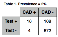
Unfortunately, this recommendation does not take into account the potential downsides to such confirmatory testing in low-risk patients, and does not consider the impact of the test threshold on clinical decision making. Potential downsides include both monetary issues (cost of the test, prolonged hospitalization, time off work) and false positive risk. A simple exercise can demonstrate the high theoretical incidence of false positive stress testing in low risk patients. Using reported accuracy measures for treadmill stress echocardiography (positive LR 7.94 and negative LR 0.19, Banerjee 2012), we can draw 2X2 tables for a hypothetical group of 1000 patients with a prevalence of coronary artery disease (CAD) of 5% (Table 1). We see that of the 50 patients with CAD, 41 will have a positive stress test, while 9 will have a false negative test. Equally as important, we see that 100 patients without CAD will have a false positive stress test. Over two-thirds of patients a with a positive stress test do not have CAD in this example. These 100 patients will likely be subjected to further testing, many undergoing invasive coronary angiography as a result. If we instead take a group of patients with a 2% prevalence of disease, we see that the false-positive risk increases, and potential for harm (relative to the potential for benefit) increases significantly (Table 2).
Studies have demonstrated this high false positive rate in both “low risk” and “intermediate risk” patient populations. This high risk of false positive stress testing is underscored by its recent inclusion as part of the American College of Cardiology contribution to the Choosing Wisely campaign. While these recommendations were targeted at screening in asymptomatic patients rather than symptomatic ED patients, the same risks of “invasive procedures” and “excess radiation exposure” applies to any population at significantly low risk. It is therefore imperative to identify ED patients at sufficiently low risk of ACS such that further cardiac imaging is unnecessary. The use of clinical decision rules (CDRs) and accelerated diagnostic protocols (ADPs) may help solve this crisis.
The ASPECT trial looked at an ADP that consisted of a thrombolysis in myocardial infarction (TIMI) score of 0 and normal cardiac biomarkers (troponin and CK-MB) at presentation and 2 hours later. This ADP identified a population with a 0.9% risk of a major adverse cardiovascular event (MACE) at 30 days, with a sensitivity of 99.3% and a negative predictive value (NPV) of 99.1%. There were 3 patients in the study with a false negative ADP, 2 of whom required coronary stenting and one of whom underwent radiofrequency ablation for a 30-minute episode of stable ventricular tachycardia; none died or had serious morbidity. A follow-up study (Aldous 2012) looked at several CDRs and ADPs in patients from one institution in the ASPECT trial. Three ADPs were found to be superior, including the 2-hour TIMI (sensitivity 99.2%, NPV 98.1%), an ADP reported by Hess et al (sensitivity 99.7%, NPV 98.9%), and an ADP reported by Christensen et al (sensitivity 99.4%, NPV 93.8%). Of these, the 2-hour TIMI had the highest specificity (23.8%) while the ADP by Christensen had the lowest (4%), and would likely have limited clinical utility as a result.
An evaluation of the HEART score from Wake Forest Baptist Medical Centre (Mahler 2011) demonstrated a high NPV (99.4%). It’s poor sensitivity (58.3%) was due to the inclusion of only low-risk patients in the cohort (those with a TIMI score < 2 and low clinical suspicion of ACS). Combination of the HEART score with a negative 4 to 6-hour troponin increased both the sensitivity and NPV to 100%. Unfortunately, the study protocol required the use of two CDRs (TIMI and HEART) for inclusion, making application to everyday practice difficult. Poor methodology also makes the study’s results unreliable.
Beyond ADPs and CDRs, Jeff Kline has looked at the use of attribute matching to provide pre-test probabilities for patients at risk for ACS. A recent randomized controlled trial assessed the impact of providing these pre-test probabilities to patients and clinicians in the ED. While this practice reduced the rate of negative testing associated with significant radiation exposure (> 5 mSv), this effect was not the result of reduced provocative testing, but rather a shift from radiologic testing to non-radiologic testing in the intervention group. It is unclear why attribute matching would cause such a shift in the type of tests ordered by the clinician. Further benefit should be shown prior to widespread implementation of the tool in clinical practice, especially given its proprietary nature and the associated cost.
Consensus:
While most present agreed that reducing stress testing in patients at very low risk of adverse outcomes would likely benefit both patients and society, the quantification of what constituted significantly low risk was variable, ranging from 0% up to 5%. Medicolegal risk was cited as the primary concern when discharging low-risk patients without provocative testing. Ongoing research into shared decision making may help remove some of these barriers, decrease healthcare utilization, reduce the risk of false-positive stress testing, and improve patient care.
Intranasal Naloxone in Acute Opiate Overdose
Aug 01, 2013
Show notes
Journal Club Podcast #4: July 2013
Emergency Physician and Toxicologist Evan Schwarz sits down with me to discuss intranasal naloxone administration...
It’s a typical busy shift in TCC. Your attending is busy dealing with a 75 year old with transient hypotension and syncope that was upgraded to a level 1 trauma due to his forehead abrasion when a car pulls up to the trauma bay, dumps out a 24 year old male who is apneic, and drives away. As you get him into a room, you notice the pinpoint pupils, track marks covering all his extremities, and the burned spoon that falls out of his pocket. You order naloxone but unfortunately no one is able to insert a peripheral IV.
Using your expert ultrasound skills, you place a 22 gauge IV in his thumb and revive him. You spot your attending as you leave the room and let him know about your nice save. Instead of the “great job!” that you are expecting, he just asks “well why didn’t you use a different route to administer the narcan?” Feeling discouraged, you make your way back to EM1 and spot Dr. Halcomb and Dr. Mullins in the hall. You go to ask them their opinion about giving narcan by a different route but notice that they are having a lively conversation about the Krebs cycle. Deciding you don’t want any part of that, you go back to Dr. Cohn’s office to do your own literature search.
PICO Question:
Population: Adult patients with acute opiate overdose with respiratory depression and/or altered mental status
Intervention: Intranasal naloxone
Comparison: Intravenous, intramuscular, or subcutaneous naloxone
Outcome: Time from first contact or drug administration to significantly increased respiratory rate or improved mentation, need for rescue dose of naloxone, healthcare cost, duration of emergency department (ED) incidence of needlestick injury by providers.
Search Strategy:
After quickly searching Google and pulling up a variety of entertaining pictures about heroin intoxication, you decide that Pubmed might be more useful. You enter a number of search terms including “narcan,” “naloxone,” “opiate,” “heroin,” “intranasal,” “intramuscular,” and “subcutaneous.” You eventually simplify your search, entering “intranasal naloxone,” (http://tinyurl.com/nztrfdb)resulting in 46 articles. You scan these and come up with the following 4 articles.
Bottom Line:
Opiate overdose remains a significant health problem in the US and elsewhere, with increasing overall rates of overdose and death related to overdose observed (Hall 1999, Preti 2002, Shah 2008, Bryant 2004). Naloxone remains the second most commonly administered antidote in the US (Wiegand 2012). While naloxone is typically administered either by intravenous (IV) or intramuscular (IM) injection, other potential routes of administration have been proposed, including nebulization (Baumann 2013, Tataris 2013), endotracheal injection (Tandberg 1982), and subcutaneous (SQ) injection (Wanger 1998). High rates of needlestick injuries have led some to propose avoiding IV, IM, and SQ naloxone administration, with many favoring intranasal (IN) administration as first-line treatment in the management of opiate overdose.
In addition to reducing the risk of percutaneous needlesticks among healthcare workers, IN naloxone administration has other potential benefits over other routes. Theoretically, naloxone should have 100% bioavailability through the nasal mucosa with a similar onset of action and similar half-life to IV administration. While rat studies have demonstrated such pharmacodynamics, a single human volunteer study showed poor bioavailability (4% vs. 35%) for the IN vs. the IV route (Dowling 2008); however, this study used a more dilute solution of naloxone than typically recommended (2 mg in 5 mL) and did not address the impact of IN naloxone in actual opiate overdose.
Two retrospective chart reviews were identified assessing the utility of IN versus IV naloxone in the prehospital setting. The study by Robertson demonstrated similar rates of clinical response (66% and 56% in the IN and IV groups, respectively) and similar mean time from patient contact to clinical response (20.3 vs. 20.7 minutes in the IN and IV groups, respectively). The study by Merlin demonstrated similar median increases in respiratory rate (RR) (4 vs. 6 breaths/minute) and Glasgow Coma Scale (GCS) (3 vs. 4) between the IN and IV groups.
Unfortunately, these studies suffered from several major methodological flaws. These were both retrospective chart reviews using prehospital records. Neither study reported on appropriate chart review methods, including abstractor training, use of standardized abstraction forms, blinding of abstractors to the study hypothesis, or assessment of interrater reliability. Additionally, there was no standardized or independent method used to assess outcomes such as respiratory rate, time elapsed, or the need for additional doses of naloxone, and no interrater reliability was assessed for these measurements. The study by Merlin assessed changes in RR and GCS over the course of the EMS encounter, but did not assess the duration of time over which these changes occurred. While the study results suggest that IN naloxone is as effective as IV naloxone, the poor methodological quality of the studies brings this conclusion into question.
Two randomized controlled trials were also identified, assessing IN versus IM naloxone in acute opiate overdose in the state of Victoria, Australia. The two studies were conducted by the same group, 4 years apart. In the first study, the IM group required less time from naloxone administration to achieve a respiratory rate of 10 breaths per minute than the IN group (mean 6 minutes vs. 8 minutes, p = 0.006), and was more likely to achieve a respiratory rate of 10 or more by 8 minutes after naloxone administration (82% vs. 63%, p = 0.0163). The IN group did have a lower rate of agitation/irritation (2% vs. 13%), and there was no statistically significant difference in the rates of minor adverse events. There was no difference in the need for rescue naloxone. The second study demonstrated similar rates of adequate response within 10 minutes of initial naloxone administration between the IN group and IM group (72.3% vs. 77.5%), as well as similar mean response times (8.0 minutes vs. 7.9 minutes). More patients in the IN group required rescue naloxone (18.1%) compared to the IM group (4.5%). There was no difference between the two groups with respect to minor adverse events, hospitalization rates, agitation/violence, nausea/vomiting, or headache.
While these studies were randomized, the providers, patients, and outcome assessors were not blinded to treatment group; no sham or placebo treatments were given. As in the previous two studies, there was no standardized, objective, independent means of measuring time or respiratory rate, or of determining the need for rescue naloxone. The difference in outcomes between these two studies was addressed by the authors, who conceded that the first study used a diluted solution of naloxone in the IN group, administering 5 mL via atomizer instead of the standard 1 mL in order to deliver 2 mg of naloxone (Wolfe 2004). As previously stated, this may lead to decreased bioavailability, likely due to the inability of the nasal mucosa to absorb such a large volume leading to a significant amount of medication being swallowed.
Consensus
Given the similar rates of minor adverse events in these four studies, and the low rate of major adverse events (one seizure in a patient receiving IM naloxone in the Kerr study), all three routes seem reasonably safe. Despite all of the authors' reported concerns over needlestick injury in healthcare workers administering IV and IM naloxone, there were no reported needlestick injuries in any of the studies. The current evidence suggests that IN naloxone is both safe and effective, though may require a rescue dose of naloxone be administered in many cases. The consensus among our group was that few would use IN naloxone as first-line therapy for opiate overdose, but that it would be viable alternative in certain circumstances (e.g. patients with difficult IV access, patients requiring extraction from vehicles).
In addition to healthcare provider administered IN naloxone, peer distribution of IN naloxone has been studies in the US (Doe-Simkins 2009). A survey of Australian heron users revealed strong support for the practice, and some groups have pushed for further research in this area (Lenton 2000, Lenton 2009) as well as the UK (Wright 2006). Potential problems with peer distribution include the medico-legal risk of prescribing medications that will most likely be administered to people other than those for whom they were prescribed, the cost of distributing a sufficient supply of naloxone to have significant impact on overdose morbidity and mortality, and failure of bystanders to contact EMS after administering naloxone, potentially leading to increased morbidity and mortality in cases of naloxone failure or overdose recrudescence (Darke 1997). Further research to address these concerns will likely be necessary before widespread implementation.
Cardiac Ultrasound in Cardiac Arrest
Jul 08, 2013
Show notes
Journal Club Podcast #3
Ultrasound expert Dan Theodoro and I discuss the use of cardiac ultrasound in predicting outcomes in cardiac arrest...
A shift in TCC yields a now all too familiar dilemma. At peak hours you must manage a patient who has sepsis and requires a central line to follow SCVO2. Your other patient is an asthmatic who, after overstaying the allotted 23 hours in your observation unit by 4 hours, can’t speak in full sentences despite a tripod stance and BiPAP. A level 1 hypotensive trauma patient with a pelvic fracture awaits VIR. The nurses hand you an EKG and you shriek because, without even looking down, the machine is reading ACUTE MI and this time, it’s right. As you groan thinking it can’t get worse than this, it does... EMS brings in a 45 year old healthy appearing patient in full cardiac arrest, down an unknown number of minutes on the scene. It’s unclear, but EMS says bystanders were there without an AED but providing compressions for most of time your patient was down. Just to make it worse, EMS is walking in your patient’s 5 and 7 year old children to the social work office while their parent goes into TCC-1, Left.
“You don’t have time for this,” whispers a little voice in your head that on this occasion might be right. As the nurses help move the patient and place monitor leads on the chest, you confirm ETT placement and find out EMS delivered 3 rounds of epinephrine without good effect. You ask to hold compressions but feel no pulse despite narrow complexes going across the monitor at a rate of about 80. Compressions continue, you perform bilateral chest decompression, administer another round of epinephrine, calcium gluconate, and bicarb, and wait 2 minutes while compressions continue. Compressions are held but you feel no pulse (other than your heart pounding as the nurse tells you the AMI looks grey and your septic patient is hypotensive); the monitor again shows PEA. The team, looking desperate, wants you to “call it.” You pull out your ultrasound hoping that might show you a cardiac effusion you can drain and story you can tell for the ages but your patient is not so lucky. All you see is standstill. Total resuscitation time gone by: 19 minutes. You ask yourself, “Can I call it? Do I have enough information to tell those kids their parent is gone and there’s nothing I can do about it?”
Time stops! Chris Carpenter bounds in, computer in hand, followed by Brian Cohn. They both have on ninja outfits. Chris’ is all black with the words “Knowledge” and “Power” embroidered down the sleeves. Brian’s is white with a green belt. Carpenter quickly assesses what your PICO should be but hesitates a second to get it going as his mentor’s words echo in his head, “He who spreads himself too thin ends up with margarine.” Reluctantly (or maybe not so reluctantly), he hands the computer off to Brian Cohn who, trembling in the presence of EBM greatness but newly armed with McDaddy EBM knowledge from McMaster’s EBM course, puts his head down and grunts “Let’s get it on!”
PICO Question:
Population: ED patients who present in cardiac arrest
Intervention: Bedside ultrasound
Comparison: Clinical gestalt, end-tidal CO2
Outcome: Return of spontaneous circulation, survival to hospital admission, survival to hospital discharge, meaningful neurologic recovery Search Strategy:
Out of nowhere Brian inputs (((heart arrest OR cardiopulmonary resuscitation OR cardio-pulmonary resuscitation OR CPR OR advanced cardiac life support OR cardiac arrest OR asystole) AND (echocardiography OR ultrasonography OR echocardi* OR echo OR cardiac echo OR cardiac ultrasound OR cardiac ultrasonography OR TTE OR transthoracic echocardiography OR transthoracic echocardiogram OR trans-thoracic echocardiogram OR trans‐thoracic echocardiography OR ultrasound OR sonogram) AND ((incidence[MeSH:noexp] OR mortality[MeSH] OR follow up studies[MeSH:noexp] OR prognos*[TextWord] OR predict*[TextWord] OR course*[TextWord] or death[TextWord]) OR (predict*[tiab] OR predictive value of tests[mh] OR scor*[tiab] OR observ*[tiab] OR observer variation[mh])))) into pubmed. The search yields several articles from which you pick these 4...
Bottom Line:
Out-of-hospital cardiac arrest remains a leading mechanism of death in the United States, with an estimated incidence of 300,000 events per year (McNally 2011). Overall survival has remained stable at approximately 8% since the 1950s (Sasson 2010) despite initiatives to improve survival rates (improved bystander CPR, public use of automated external defibrillators). Given the very low likelihood of survival in patients presenting to the ED without a pulse (0.9% in one large database), efforts have been made to determine those in whom ongoing resuscitation is futile.
While some recommend bedside cardiac ultrasound as a diagnostic tool in cardiac arrest to aid in detection of reversible conditions such as severe hypovolemia, pericardial tamponade, and massive pulmonary embolism (Hernandez 2008), there has also been widespread use to evaluate for the absence of cardiac activity, with the results frequently affecting the duration of resuscitation efforts. In one survey of graduates from the LA County/USC Medical Center residency program, 68% reported using ultrasound during cardiac arrest, and 91% of these reported using the results in deciding when to terminate resuscitation efforts (Schoenberger 2007). It is important, however, to review the evidence surrounding this practice.
We identified three primary studies (Blavais 2001, Salen 2001, Aichinger 2012) and one meta-analysis (Blyth 2012) dealing with this subject. In patients in cardiac arrest with no cardiac activity on ultrasound, all of the papers revealed low rates of survival to hospital admission, ranging from 0% to 3.1%. While no clearly identified survival threshold exists above which resuscitation should be continued, given the severity of the outcomes a low threshold should be used. The pooled survival to hospital admission rate in the meta-analysis of 2.4% (95% CI 1.3-4.5%) would indicate that cardiac ultrasound should not be used alone to to determine when further attempts at resuscitation are futile. Unfortunately, the meta-analysis and two of the studies did not assess survival beyond admission or neurologically intact survival. The one study to assess survival to hospital discharge (Aichinger 2012) found that none of the 31 patients with cardiac standstill survived to discharge, however the width of the 95% confidence interval (0%-11%) prevents us from safely applying these results to clinical practice.
There was consensus among those present that these studies do not support stopping resuscitation efforts in cardiac arrest patients based on the absence of cardiac activity on ultrasound. This decision was based on both the size of the studies (and resulting width of the confidence intervals), and the lack of assessment of long-term outcomes (beyond hospital admission/discharge). Many felt that future studies should exclude patients in ventricular fibrillation or ventricular tachycardia, as standard care would dictate ongoing resuscitation in these patients. Future studies will also need to beware the potential adverse consequences of pausing CPR for ultrasound performance, making sure to time this with pulse checks.
One of the issues in these studies was the use of relatively short-term outcomes. The meta-analysis and two of the primary studies evaluated survival to hospital admission as the primary outcome. More clinically important outcomes may include survival to hospital discharge, 30-day or one-year survival, and neurologically intact survival assessed by modified Rankin Scale or CPC score. A conference of the Research Working Group of the American Heart Association Emergency Cardiovascular Care Committee to discuss appropriate outcomes in resuscitation research demonstrated the difficulties in such studies. There was no consensus on a single appropriate outcome, and conference participants were unable to agree on the ideal outcome measure when confronted with 4 hypothetical cases. There was consensus that large trials designed to have a major impact should use longer-term endpoints at least 90 days out coupled with some neurological and quality-of-life assessment.
There are currently no registered trials addressing long-term outcomes of cardiac arrest patients with cardiac standstill on ultrasound. The Reason 1 trial is currently recruiting patients to assess survival to hospital discharge, and the investigators plan to enroll 1000 patients. This would make it the largest study to date on this subject. Based on the expected results of this trial, the investigators plan on gaining funding to conduct a similar trial that will look at long-term outcomes in these patients.
The INTERACT2 Trial- Blood Pressure Reduction in Spontaneous Intracerebral Hemorrhage
Jun 24, 2013
Show notes
Patients with hemorrhagic stroke, or intracerebral hemorrhage (ICH), are common in emergency medicine and frequently present to the emergency department with significantly elevated blood pressures requiring urgent treatment. The correct goal blood pressure is debatable. On one hand there is concern that the hydrostatic effects of elevated blood pressure could contribute to expansion of the hematoma, increased edema, or rebleeding. On the other hand, overcorrection of the blood pressure could lead to decreased cerebral perfusion in the setting of autoregulation.
Currently, the AHA/American Stroke Association recommendations for the management of blood pressure in spontaneous (atraumatic) ICH are as follows:
1) If SBP is > 200 mmHg or MAP is >150 mmHg, then consider aggressive reduction of BP with continuous intravenous infusion.
2) If SBP is >180 mmHg or MAP is >130mm Hg and there is the possibility of elevated ICP, then consider monitoring ICP and reducing BP using intermittent or continuous intravenous medications while maintaining a cerebral perfusion pressure >60 mmHg.
3) If SBP is >180 mmHg or MAP is >130 mmHg and there is no evidence of elevated ICP, then consider a modest reduction of BP (e.g. MAP of 110 mmHg or target BP of 160/90 mmHg) using intermittent or continuous intravenous medications to control BP.
4) In patients presenting with a systolic BP of 150 to 220 mmHg, acute lowering of systolic BP to 140 mm Hg is probably safe
Note that these recommendations are Class C, based partly on the INTERACT pilot study, which looked at 440 patients and compared goal SBP of 140 mmHG with more modest reductions to 180 mmHG. They found a trend toward reduced hematoma growth with no increase in neurologic deterioration or differences in other clinical outcomes (disability, quality of life). The study was underpowered to detect significant differences in any of the outcomes.
Methods:
INTERACT2 was an international, multicenter, prospective, randomized trial. The study was open-treatment by necessity; patients and providers could not be blinded to group allocation due to the nature of the study. They were blinded to the end-point of the trial, and outcome assessors were blinded to group allocation. Patients were excluded if they had a definite contraindication to BP-lowering treatment, if treatment could not be initiated within 6 hours of ICH, if there was a structural cause for the ICH, if they had a GCS of 3-5 (“deep coma”), if they had a massive hematoma with poor prognosis, or if early surgical evacuation of the hematoma was planned.
Patients were randomized to two groups: 1) in the intensive-treatment group, IV and oral antihypertensive were initiated by protocols based on the local availability of agents, with the goal of achieving a SBP < 140 mmHg within one hour of randomization and maintaining this level for 7 days; 2) in the standard-treatment group, BP-lowering treatment was initiated if the SBP was > 180 mmHg, with no lower level stipulated. All patients were started on oral antihypertensives within 7 days or hospital discharge. Patients were followed-up in person of by telephone at 28 days and 90 days by local staff blinded to treatment group. Patients were analyzed by the intention to treat principle.
The primary outcome was the proportion of patients with a poor outcome: death or severe disability (a score of 3-5 on the modified Rankin scale at 90 days after randomization). Initially, the key secondary outcome was death or severe disability in patients in whom treatment was initiated within 4 hours of onset of ICH. This was changed prior to data analysis to physical function across all 7 levels of the modified Rankin scale using ordinal analysis. Other secondary outcomes included: all-cause mortality; cause-specific mortality; health related quality of life; duration of hospitalization; residence at a residential care facility at 90 days; poor outcomes at 7 and 28 days; and serious adverse events (neurologic deterioration or severe hypotension). They also assessed change in hematoma size in a subset of patients who underwent repeat CT or MRI at 24 hours post-ICH.
Between October 2008 and August 2012, 2839 patients were enrolled at 144 hospitals in 21 countries; 1403 were assigned to intensive-treatment and 1436 were assigned to standard treatment. Patients were similar with respect to known risk factors…with the exception of warfarin use (3.6% in the intensive-treatment group, 2.2% in the standard-treatment group, p = 0.025). Approximately 68% of all patients were recruited from Chinese hospitals. In terms of treatment, patients in the intensive-treatment group received antihypertensives more quickly and had more rapid lowering of BP than those in the standard-treatment group. Of note, patients in the intensive-treatment group were more likely to have care withdrawn than those in the standard-treatment group (5.4% vs. 3.3%, p = 0.005). The primary outcome was assessed in 98.5% of intensive-treatment patients and 98.3% of standard-treatment patients.
Results:
For the primary outcome, poor outcome at 90 days (death or score of 3-5 on the modified Rankin scale), there was no difference between the intensive-treatment (52%) and standard-treatment (55.6%) groups (OR 0.87; 95% CI 0.75-1.01). Ordinal analysis showed a favorable shift in the distribution of scores on the modified Rankin scale for patients in the intensive-treatment group (pooled OR for a shift to higher score of 0.87, 95% CI 0.77-1.00). The rate of death from any cause was similar for the intensive-treatment and standard treatment group (11.9% vs. 12.0% (OR 0.99; 95% CI 0.79-1.25) as was the percentage of deaths directly attributed to the ICH, duration of hospitalization, and rates of serious adverse events.
Patients in the intensive-treatment group reported better quality of life at 90 days using the European QOL 5-dimension questionnaire which assesses mobility, self-care, usual activities, pain or discomfort, and anxiety or depression (each graded on a 3-level scale as either no problems, moderate problems, or extreme problems) than those in the standard-treatment group: mean utility score 0.6 vs. 0.55. p = 0.002.
For patients who underwent repeat brain imaging at 24 hours (35.1% of the intensive-treatment group and 33.1% of the standard-treatment group) there was no significant difference in the mean hematoma growth: relative difference 4.5% (95% CI -3.1% to 12.7%), absolute difference 1.4 mL (95% CI -0.6 mL to 3.4 mL).
Commentary:
The authors note that while intensive lowering of BP in ICH did not result in a significant reduction in the primary outcome, there were better functional outcomes when an ordinal analysis of the primary outcome was undertaken. The ordinal analysis showed a very slight benefit with intensive lowering of BP, with a 95% CI that just reaches 1.00 (and a p-value of 0.04). Keep in mind that the authors chose to perform ordinal analysis after the data had been collected. One should always beware when investigators change the study in some way after data collection has been initiated. In this case, the authors’ reasoning was that ordinal analysis began gaining acceptance in clinical trials partway through the course of the trial. Also keep in mind that this is the secondary outcome, not the primary outcome, which was selected a priori. For the other secondary outcome of quality of life, there was a slight improvement with intensive lowering of BP with a mean EQ-5D score of 0.6 vs. 0.55. The clinical significance of this difference is unfortunately difficult to interpret. If my overall score is 0.6, how much better off am I than someone with a score of 0.55?
Issues of external validity have been raised as well, given that the majority of patients in the study (68%) were recruited from Chinese institutions. We have to ask two questions: 1) are Chinese patients with ICH somehow different than patients in our own institutions? and 2) is the treatment of ICH in Chinese institutions different than treatment at our institutions? Given that the majority of patients with spontaneous ICH are treated with supportive care, and that the indications for invasive procedures (ICP monitoring, craniotomy/craniectomy) are likely the same at any large hospital with neurosurgical capability, I would suspect that these results are likely valid in the US. Additional studies at US institutions may help elucidate this further.
Bottom Line:
The authors conclude: “early intensive lowering or BP in this patient population is safe,” which is in keeping with the AHA/ASA guidelines, which state “In patients presenting with a systolic BP of 150 to 220 mmHg, acute lowering of systolic BP to 140 mm Hg is probably safe.” I tend to agree with these statements. This study alone should by no means make intensive lowering of BP in ICH standard of care, as there is no clear benefit.
EBM Teaching Point:
The authors in this study used an ordinal analysis of the modified Rankin score. The Rankin score is an example of a type of categorical data known as ordinal data, in which numerical scores are arbitrarily assigned to levels of measurement. The assigned number itself has no meaning, other than to provide a scale for reference. Examples include pain scores (0 out of 10 means no pain, 10 out 10 is severe pain). Classically, studies that look at outcomes based on ordinal data have applied a cutoff, thus turning a scale with multiple levels into a binary outcome. Instead of worrying about whether you’re a 1, 2, 3, etc., we only care about whether you’re above or below the cutoff. We can then compare the proportions above or below the cutoff in 2 groups and assign p-values or chi-square values. Ordinal analysis on the other hand uses more complex statistical calculations to assess shifts across the entire spectrum of the scale. This has been shown to have higher statistical power than using binary outcomes alone.

 Our TOPPODCAST Picks
 Stay Connected